Historic Surgeon’s First Successful Cross-Circulation Surgery Patient Donates Body to Visible Heart Lab
A special donation to the Visible Heart Laboratories at the University of Minnesota Medical School will inform surgeons at the U and around the world about the effects of one of the world’s first open-heart surgeries performed by C. Walton Lillehei, PhD, MD, a professor in the Department of Surgery from 1951 to 1967.
Paul A. Iaizzo, PhD, professor, and Tinen Iles, PhD, assistant professor, who are both investigators within the Visible Heart Laboratories, performed an educational dissection of the patient who received the world’s first successful open-heart surgery using cross-circulation in 1954 after he passed away and donated his body to the lab in 2018. In January, the team published their findings from the educational dissection in the journal, Annals of Thoracic Surgery.
Cross-Circulation: World’s First Open-Heart Surgeries
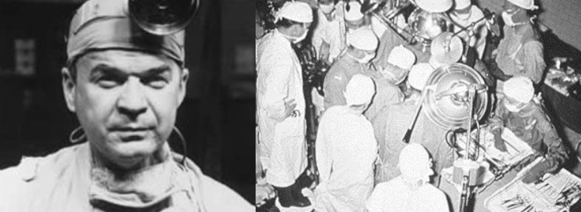
The University made a world-renowned impact in cardiac surgery by performing the world’s first open-heart surgery using cross-circulation. These were the first open-heart surgeries in the world where they had direct visualization to stop the heart, pull the blood out, see the defect and then make the surgical repair. Dr. Lillehei and his team used cross-circulation, a procedure where one individual provided oxygenated blood to the other person who was undergoing open-heart surgery. This method, at the time, was an innovative way to keep the cardiac patient’s brain and vital organs working while doctors stopped blood flow to the heart for the repair. In most cases, the surgery was performed on a child with the parent supporting the oxygenated blood.
These procedures were established in 1954 and were used in some cases for patients who needed a large ventricular defect (VSD) repair. VSD is a defect in which someone is born with and is one of the most common congenital heart diseases; it’s a hole in the ventricle between the left and right side of the heart, which is an issue because the left ventricle is then pushing partially deoxygenated blood out to the rest of the body when it should be fully oxygenated. Without a procedure to fix the hole, the patient would eventually become very incompacitated and may even die.
Dr. Lillehei’s first successful cross-circulation patient in April 1954 was three years old, and his 25-year-old father provided him with the oxygenated blood needed during the procedure. His family, knowing the important history of the patient’s surgery, donated his body to the University’s Bequest Program and then to Visible Heart Laboratories for research and educational purposes.
“These procedures and this patient really moved the whole field of congenital heart repair forward,” Dr. Iaizzo said.
VSD repairs are still performed at the University today, mostly led by Massimo Griselli, MD, MS, FRCS, professor in the Department of Surgery. Though cross-circulation is no longer used because of new technologies, Dr. Griselli says these early procedures laid a foundation for today’s open-heart surgeries.
“Dr. Lillehei completely revolutionized the world of cardiatric surgery through his cross-circulation procedure. Before there was nothing else available for patients, they were just dying,” Dr. Griselli said. “Since then, we’ve been able to make it an almost perfect type of approach with VSD. This has been an extraordinary achievement made at the University of Minnesota, and it’s truly something amazing what they have done.”
Using History to Advance the Future of Medicine
From this patient’s donation, Drs. Iaizzo and Iles and Mikayle Holm, a PhD student, were able to conduct research through CT imaging of the body that enabled them to create an entire reconstruction of the patient’s heart and circulatory system. Dr. Iles said cameras inside of the heart made it possible to view scars from previous surgeries.
“I think the most notable part for me was seeing the scar tissue and that the heart was so enlarged. It took up most of the space in his chest,” Dr. Iles said. “It’s remarkable he lived such a long life.”
Additionally, when Dr. Lillehei passed away in 1999, he donated archives from his office and home to the University. Drs. Iaizzo and Iles visited these files at the Anderson Library and watched actual video footage from Dr. Lillehei’s surgeries to receive other relevant information about the patient.
“It was all really fascinating to look at,” Dr. Iaizzo said.
About six months after receiving the patient’s body, his family reached out to the Visible Heart Laboratories and asked to take a visit. Drs. Iaizzo and Iles provided historic presentations to the patient’s family. They also showed them the unique computational models created of his vasculature and heart, including a 3D-printed heart model that was painted by Holm to look like a real, human heart.
“They were so moved by it all that they asked if they could have the 3D print to show the rest of the family, so we gave them one,” Dr. Iaizzo said.