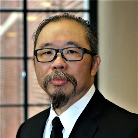
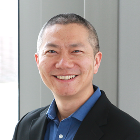
Faculty
Department Chair
Charles Dietz, MD
Chair and Associate Professor of Radiology
dietz004@umn.edu
Administrative Contact
Laura Geffre-Rick
geffr019@umn.edu
612-301-6040
Education
Fellowships, Residencies, and Visiting Engagements
Contact
Address
420 Delaware Street, SE, MMC 292, Minneapolis, MN 55455Administrative Contact
Chelsey Grandstrand
Administrative Coordinator
Email: grandscr@umn.edu
Phone: (612) 612-2742
Fax: (612) 626-5505

Bio
Dr. Mangia is a Professor in the Department of Radiology at the University of Minnesota. She got her master degree in Physics (1999), the Ph.D. in Biophysics (2003) and the title of "Specialist in Medical Physics" (2005) at the University of Rome "La Sapienza" (Italy). Since 2004 she has been working at the Center for Magnetic Resonance Research (CMRR).
Research Summary
Functional studies of human brain metabolism under increased or decreased neuronal activity
Neurovascular coupling and neuro-glia interactions
Oxidative and non-oxidative brain metabolism; glucose and lactate metabolism
Novel MR contrasts based on rotating frame relaxation measurements, magnetization transfer and chemical exchange
Applications of novel MRI/MRS protocols for studies of neurological diseases as Parkinson's disease, schizophrenia, multiple sclerosis Measurements of blood flow with arterial spin labeling methods, and their application to diseases as diabetes
Proton NMR Spectroscopy at high fields
Resting state fMRI, especially in relation to brain metabolism
Education
Fellowships, Residencies, and Visiting Engagements
Professional Memberships
Contact
Address
2021 6th Street SE, Minneapolis, MN 55455
Education
Fellowships, Residencies, and Visiting Engagements
Licensures and Certifications
Professional Memberships
Contact
Address
420 Delaware Street, SE, MMC 292, Minneapolis, MN 55455Administrative Contact
Chelsey Grandstrand
Administrative Coordinator
Email: grandscr@umn.edu
Phone: (612) 612-2742
Fax: (612) 626-5505
Bio
Małgorzata (Gosia) Marjańska is a professor in the Department of Radiology. Dr. Marjańska received a B.S. in chemistry with minor in mathematics from Loyola University of Chicago and a Ph.D. in chemistry from the University of California at Berkeley. She joined the Center for Magnetic Resonance Research (CMRR) in 2002 as a post-doctoral fellow working with Prof. Kamil Ugurbil and subsequently became a research assistant professor two years later, and assistant professor in 2013. In 2015, she was promoted to an associate professor and in 2020 to a professor.
Dr. Marjańska first encountered magnetic resonance while working with Prof. Keith Jameson at Loyola University of Chicago, first learning and then training other undergraduates to operate a 300 MHz Varian system. During the summer after her 3rd year of undergraduate studies, she worked at Bell Laboratories with Dr. Peter Mirau on solid state NMR characterization of polymer films. In order to follow her interest in NMR, she decided to pursue a Ph.D. at the University of California at Berkeley in the group of Prof. Alexander Pines. Her thesis titled "Quantum Logic Gates, Local Field, Selective Excitation and Structural Studies of Dipolar Couplings in Oriented Molecules" was focused on NMR as a technique to study molecules dissolved in liquid crystals.
Research Summary
Dr. Marjańska is interested in developing MRS techniques for humans and rodents over a wide range of field strengths (3 T up to 16.4 T) and applications of those methods to study various diseases including Alzheimer’s disease, dystonia, depression, and brain tumors. Dr. Marjańska has been interested in 1H and heteronuclear MRS (13C and 2H), diffusion-weighted MRS and relaxometry. The long-term goal of her research is to develop and assess robust, non-invasive and repeatable MRS methods for discovery of quantitative biomarkers for clinical research and practice.
Education
Fellowships, Residencies, and Visiting Engagements
Honors and Recognition
Professional Memberships
Selected Publications
Contact
Address
2021 6th Street SE, Minneapolis, MN 55455
Licensures and Certifications
Contact
Address
420 Delaware Street, SE, MMC 292, Minneapolis, MN 55455Administrative Contact
Bibi Husain
Administrative Coordinator
Email: husai002@umn.edu
Phone: (612) 626-5566
Fax: (612) 626-5505
Bio
Dr. Metzger received his B.S. from the University of Pennsylvania in 1992 and his Ph.D. from the Department of Biomedical Engineering at the University of Minnesota in 1997. His graduate research, conducted under the mentorship of Dr. Xiaoping Hu, focused on novel magnetic resonance chemical shift imaging techniques. After his graduate studies, Dr. Metzger accepted a position with Philips Medical Systems as a clinical scientist at the University of Texas Southwestern Medical Center in Dallas. In this position, he expanded his knowledge in clinical research with specific work on breast, kidney, cardiac and brain MRI applications. In his final two years of his eight year tenure with Philips, he worked at NIH as a senior clinical scientist focusing on diagnostic prostate imaging and MRI guided prostate interventions. With a desire to return to academia, he accepted a position at the University of Minnesota in 2005 with a joint appointment in the departments of Radiology and Urologic Surgery.
Research Summary
Prostate Cancer
Magnetic Resonance Imaging
Ultra-high field MRI
Magnetic resonance imaging (MRI) represents a promising method to determine the clinical significance of prostate cancer. Most men diagnosed with prostate cancer have small tumors with low inherent biological propensity for invasive growth and metastasis. These patients may be best treated through observation, or limited therapies. To the contrary, some patients have extensive, biologically aggressive tumors best treated by prostatectomy. Unfortunately, current diagnostics methods, short of pathologic examination of prostatectomy specimens, cannot reliably determine disease extent (volume and spread outside the prostate) and biological aggressiveness.
Dr. Metzger's lab is investigating the potential of magnetic resonance imaging and spectroscopy to non-invasively determine the extent and aggressiveness of prostate cancer in clinical studies. This information would be used to improve diagnosis and staging, target therapy and monitor treatment. Initial research objectives involve the development of a multi-parametric statistical model to non-invasively determine cancer probability maps based on anatomic and functional 3 Tesla MRI data. This statistical model will use registered pathology sections as a gold standard of tumor extent and co-localized molecular studies as a gold standard of aggressiveness. A second major focus of Dr. Metzger's lab involves the development of novel RF coils and imaging methods necessary to make prostate imaging at ultra-high magnetic fields feasible. The increased spatial and spectral resolution of an optimized 7 Tesla prostate imaging platform will improve the ability to track small changes in prostate cancer biomarkers facilitating the study of local disease progression and treatment response.
Education
Professional Memberships
Contact
Address
2021 6th Street SE, Minneapolis, MN 55455
Research Summary
My research is focused on relaxations during radiofrequency irradiation and development of non-invasive contrast methods for Magnetic Resonance (MR) imaging and spectroscopy at high magnetic fields (3T and higher). This research effort has resulted in numerous contributions that have fundamentally impacted brain and body research. Rotating frame relaxation methods based on adiabatic pulses were first developed in our laboratory at CMRR in Minnesota. We followed this development with a large body of work (utilizing theoretical modeling and experiments) investigating different relaxation pathways in vivo at different magnetic field strengths. The novel MR contrasts and protocols developed in my group were proven to provide an excellent tool for investigation of neurodegenerative processes in Parkinson's disease as well as cancer and stroke. It has been 3 years that we applied our rotating frame relaxation methods T1 and T2 to investigate iron accumulation and neuronal loss, respectively, in substantia nigra (SN) of Parkinson's disease patients. Recently we developed rotating frame method that comprises two relaxation pathways, T1 and T2. This method provides greater sensitivity to molecular motion and is entitled Relaxation Along a Fictitious Field (RAFF). Utilization of fictitious fields in MRI is an important aspect of research in my group.
Education
Fellowships, Residencies, and Visiting Engagements
Professional Memberships
Contact
Address
2021 6th Street SE, Minneapolis, MN 55455
Bio
Dr. Steen Moeller is a Research Associate Professor in Radiology at the Center for Magnetic Resonance Research, University of Minnesota ("CMRR"). After obtaining a Master's Degree (Cand.Scient.) in Mathematics and Physics from the University of Aalborg, Denmark in 1997, Dr. Moeller conducted research on inverse problems for electrical impedance tomography and was awarded the Ph.D. degree in 2002 from the Department of Mathematical Sciences, University of Aalborg. He joined CMRR as a post-doctoral associate for Dr. Kamil Ugurbil in 2003 and focused on mathematical algorithms for accelerated image reconstructions at ultra high fields utilizing multi-channel arrays. His work was focused on the implications for functional MRI and hardware design criteria. In 2006, Dr. Moeller joined Dr Michael Garwood's group for investigating and developed mathematical tools for the SWIFT (Sweep Imaging with Fourier Transformation) technique developed at CMRR. The technologies developed were licensed by Steady State Imaging, LLC and subsequently licensed by General Electric. Since 2010, Dr. Moeller has been involved in the technology development for the Human Connectome Project and has developed robust mathematical techniques for fast image reconstruction.
Research Summary
Mathematical techniques for accelerated image reconstruction in MRI
Quantitative and qualitative techniques for image quality evaluatio
Ultra high Field Imaging
Functional MRI
Education
Professional Memberships
Contact
Address
2021 6th Street SE, Minneapolis, MN 55455
Clinical Summary
Pediatric vascular malformations
Pediatric musculoskeletal imaging
Pediatric body MRI
Pediatric molecular imaging
Education
Fellowships, Residencies, and Visiting Engagements
Licensures and Certifications
Honors and Recognition
Professional Memberships
Contact
Address
420 Delaware Street, SE, MMC 292, Minneapolis, MN 55455Administrative Contact
Chelsey Grandstrand
Administrative Coordinator
Email: grandscr@umn.edu
Phone: (612) 612-2742
Fax: (612) 626-5505
Research Summary
Advanced imaging of brain tumors
Adrenoleukodystrophy
Inherited diseases of the brain and spine
Medical Education
Integrating Technology with Health Care
Clinical Summary
Injections for pain relief
Intraarterial chemotherapy delivery for brain tumors and retinoblastoma
Imaging of metabolic and inherited disorders of children
Brain tumor imaging
Education
Fellowships, Residencies, and Visiting Engagements
Licensures and Certifications
Honors and Recognition
Professional Memberships
Contact
Address
420 Delaware Street, SE, MMC 292, Minneapolis, MN 55455Administrative Contact
Bibi Husain
Administrative Coordinator
Email: husai002@umn.edu
Phone: (612) 626-5566
Fax: (612) 626-5505

Bio
Dr. Gülin Öz is a Professor in the Department of Radiology, Center for Magnetic Resonance Research (CMRR). Following BS degrees in Physics and Chemistry and a PhD in Biochemistry, she continued with postdoctoral training at the CMRR where she later joined the faculty.
Dr. Öz uses high field, multi-nuclear magnetic resonance spectroscopy (MRS) to delineate neurochemical and metabolic alterations in neurodegenerative diseases and diabetes. She leads a program in spinocerebellar ataxias and has studied neurochemistry in Parkinson, Huntington, Alzheimer diseases and ALS. She co-led an effort by the MRS Consensus Group to provide guidelines for MRS data acquisition and analysis, quality assessment, and interpretation. She further led a multi-site Bioengineering Research Partnership (BRP) for across-platform harmonization of advanced MRS methods. She serves as PI of a Consortium to assess COVID-19 sequelae in the brain (COVID-BRAIN) and MPI of an international clinical trial readiness study (READISCA) that aims to validate MRI and MRS biomarkers in the earliest stages of neurodegeneration in spinocerebellar ataxias. Finally, she leads the MR Biomarkers Working Group of the Ataxia Global Initiative.
Selected Publications
Chandrasekaran J, Petit E, Park YW, du Montcel ST, Joers JM, Deelchand DK, Považan M, Banan G, Valabregue R, Ehses P, Faber J, Coupé P, Onyike CU, Barker PB, Schmahmann JD, Ratai EM, Subramony SH, Mareci TH, Bushara KO, Paulson H, Durr A, Klockgether T, Ashizawa T, Lenglet C, Öz G for the READISCA Consortium. Clinically meaningful Magnetic Resonance Endpoints Sensitive to Preataxic Spinocerebellar Ataxia Types 1 and 3. Ann Neurol, doi: 10.1002/ana.26573.
Deelchand DK, Henry PG, Joers JM, Auerbach EJ, Park YW, Ratai EM, Kantarci K, Öz G (2022) Plug-and-play Advanced Magnetic Resonance Spectroscopy. Magn Reson Med, 87(6):2613-2620.
Deelchand DK, Berrington A, Noeske R, Joers JM, Arani A, Gillen J, Schär M, Nielsen JF, Peltier S, Seraji-Bozorgzad N, Landheer K, Juchem C, Soher BJ, Noll DC, Kantarci K, Ratai EM, Mareci TH, Barker PB, Öz G (2021) Across-vendor standardization of semi-LASER for single voxel MRS at 3 Tesla. NMR Biomed, 34(5):e4218.
Öz G, Deelchand DK, Wijnen JP, Mlynárik V, Mekle R, Noeske R, Scheenen TWJ, Tkáč I and the Advanced Single Voxel 1H MRS Working Group (2020) Advanced single voxel 1H magnetic resonance spectroscopy techniques: Experts' consensus recommendations, NMR Biomed, e4236, DOI: 10.1002/nbm.4236.
Öz G, Harding IH, Krahe J, Reetz K (2020) MR Imaging and Spectroscopy in Degenerative Ataxias: Towards Multi-modal, Multi-site, Multi-stage Monitoring of Neurodegeneration, Curr Opin Neurol, 33(4):451-461.
Cheong I, Deelchand DK, Eberly LE, Marjańska M, Manousakis M, Guliani G, Walk D, Öz G (2019) Neurochemical Correlates of Functional Decline in Amyotrophic Lateral Sclerosis, J Neurol Neurosurg Psychiatry, 90(3):294-301.
Joers JM, Deelchand DK, LyuT, EmirUE, Hutter D, GomezCM, BusharaKO, Eberly LE, Öz G (2018) Neurochemical abnormalities in premanifest and early spinocerebellar ataxias, Ann Neurol, 83(4):816-829.
Park YW, Deelchand DK, Joers JM, Hanna B, Kantarci K, Soher BJ, Barker PB, Park HW, Öz G, Lenglet C (2018) AutoVOI: Real-time automatic prescription of volume-of-interest for single voxel spectroscopy, Magn Reson Med, 80(5):1787-1798.
Deelchand DK, Kantarci K, Öz G. (2018) Improved localization, spectral quality and repeatability with advanced MRS methodology in the clinical setting, Magn Reson Med, 79(3):1241-1250.
Öz G, Alger JR, Barker PB, Bartha R, Bizzi A, Boesch C, Bolan PJ, Brindle KM, Cudalbu C, Dincer A, Dydak U, Emir UE, Frahm J, González RG, Gruber S, Gruetter R, Gupta RK, Heerschap A, Henning A, Hetherington HP, Howe FA, Hüppi PS, Hurd RE, Kantarci K, Klomp DWJ, Kreis R, Kruiskamp MJ, Leach MO, Lin AP, Luijten PR, Marjańska M, Maudsley AA, Meyerhoff DJ, Mountford CE, Nelson SJ, Pamir MN, Pan JW, Peet AC, Poptani H, Posse S, Pouwels PJW, Ratai EM, Ross BD, Scheenen TWJ, Schuster C, Smith ICP, Soher BJ, Tkáč I, Vigneron DB, Kauppinen RA. The MRS Consensus Group. (2014) Clinical Proton MR Spectroscopy in Central Nervous System Disorders, Radiology, 270(3):658-79.
Emir UE, Tuite PJ, Öz G (2012) Elevated pontine and putamenal GABA levels in mild-moderate Parkinson disease detected by 7 tesla ¹H MRS, PLoS One, 7(1): e30918.
Emir UE, Auerbach EJ, Van De Moortele PF, Marjańska M, Ugurbil K, Terpstra M, Tkáč I, Öz G (2012) Regional neurochemical profiles in the human brain measured by 1H MRS at 7 tesla using local B1 shimming, NMR Biomed, 25: 152–160.
Öz G, Tkáč I (2011) Short-echo, single-shot, full-intensity 1H MRS for neurochemical profiling at 4T: Validation in the cerebellum and brainstem, Magn Reson Med, 65(4):901-10.
Öz G, Nelson CD, Koski DM, Henry PG, Marjańska M, Deelchand DK, Shanley R, Eberly LE, Orr HT, Clark HB (2010) Noninvasive Detection of Pre-symptomatic and Progressive Neurodegeneration in a Mouse Model of Spinocerebellar Ataxia type 1, J Neurosci, 30:3831-3838.
Öz G, Kumar A, Rao JP, Kodl CT, Chow L, Eberly LE, Seaquist ER (2009) Human Brain Glycogen Metabolism during and following Hypoglycemia, Diabetes, 58(9):1978-85.
Tkáč I, Öz G, Adriany G, Uğurbil K, Gruetter R (2009) In vivo 1H NMR spectroscopy of the human brain at high magnetic fields: Metabolite quantification at 4T vs. 7T, Magn Reson Med, 62:868–879.
Öz G, Seaquist ER, Kumar A, Criego A, Benedict LE, Rao JP, Henry PG, van de Moortele PF, Gruetter R (2007) Human Brain Glycogen Content and Metabolism: Implications on its Role in Brain Energy Metabolism, Am J Physiol Endocrinol Metab, 292(3): E946-51
Öz G, Berkich DA, Henry PG, Xu Y, LaNoue K, Hutson SM, Gruetter R (2004) Neuroglial metabolism in the awake rat brain: CO2 fixation increases with brain activity, J Neurosci, 24: 11273-11279.
Research Summary
Dr. Öz’s research interests are:
- To develop high and ultrahigh field MRI and MRS methodology for human and rodent applications.
- To establish in vivo MRI and MRS biomarkers of neurodegeneration, in parallel studies with patients and transgenic mouse models. Such biomarkers are expected to facilitate early disease detection and treatment monitoring in pre-clinical and clinical trials.
- To determine the effects of diabetes and the hypoglycemic consequences of intensive insulin therapy on brain glucose and glycogen metabolism and to elucidate pathogenic mechanisms of impaired awareness of hypoglycemia using MRS.
Education
Fellowships, Residencies, and Visiting Engagements
Honors and Recognition
Professional Memberships
Grants and Patents
Selected Grants
Patents
Contact
Address
2021 6th Street SE, Minneapolis, MN 55455
Research Summary
Pediatric/Adult brain tumor imaging
Toxic leukoencephalopathy
Non-vascular spine interventions
Spinal muscular atrophy
Molecular imaging/oncologic imaging
Cardiac PET/CT
Clinical Summary
Pediatric/Adult brain tumor imaging
Toxic leukoencephalopathy
Non-vascular spine interventions
Spinal muscular atrophy
Molecular imaging/oncologic imaging
Cardiac PET/CT
Education
Fellowships, Residencies, and Visiting Engagements
Licensures and Certifications
Professional Memberships
Contact
Address
420 Delaware Street, SE, MMC 292, Minneapolis, MN 55455Administrative Contact
Bibi Husain
Administrative Coordinator
Email: husai002@umn.edu
Phone: (612) 626-5566
Fax: (612) 626-5505

Education
Fellowships, Residencies, and Visiting Engagements
Contact
Address
420 Delaware Street, SE, MMC 292, Minneapolis, MN 55455Administrative Contact
Bibi Husain
Administrative Coordinator
Email: husai002@umn.edu
Phone: (612) 626-5566
Fax: (612) 626-5505
Bio
Dr. Patriat is an Assistant Professor in the Department of Radiology at the University of Minnesota. After receiving a B.A. in Physics and Film Studies (2010) from the University of Minnesota-Morris, Dr. Patriat earned a M.S (2012) and a PhD (2015) in Medical Physics from the University of Wisconsin. Dr. Patriat has been working on ultra-high field (e.g., 7Tesla) MRI research and its application towards the creation of patient-specific anatomical models to improve DBS targeting and efficacy, first as a Postdoctoral Researcher at CMRR (2015-2019) and then as Faculty (since 2019). Dr. Patriat’s work includes manual segmentation and machine learning applications based on direct anatomical visualization as well as structural and functional connectivity analyses.
Research Summary
Use of ultra-high field MRI technology to improve neuromodulation applications such as Deep Brain Stimulation surgery
Studying brain disorders, including movement disorders, using high-resolution resting-state functional MRI and diffusion imaging
Patient-specific brain modeling using ultra-high field MRI technology and high-resolution images
Translational research focusing on neuroimaging methods and neuroscience
Visualization and characterization of brain anatomy and connectivity networks
Licensures and Certifications
Professional Memberships
Contact
Address
2021 6th Street SE, Minneapolis, MN 55455
Bio
Dr. Pramod Pisharady is an Assistant Professor at the Center for Magnetic Resonance Research (CMRR, Department of Radiology) at the University of Minnesota Medical School. After earning his PhD in Computer Vision and Machine Learning from NUS (2012), he did postdoctoral research in the Biological Engineering department at MIT (2013-2014). Before his appointment as Assistant Professor (2022) at CMRR, he worked as a Postdoctoral Associate (2014-2017) and a Research Associate (2017-2022) at CMRR. Dr. Pisharady is a recipient of the Chan Zuckerberg Initiative (CZI) Imaging Scientist grant award (2020). His current research interest is to develop methods to bridge the information gap between macroscale and microscale imaging using multimodal imaging with diffusion MRI and microscopy, and to apply these methods to detect disease-related structural changes in neurological disorders.
Awards & Recognition
- Chan Zuckerberg Initiative (CZI) Imaging Scientist Grant Award, 2020
- Best Poster Award, Institute for Research in Statistics and its Applications (IRSA), 2017
- Travel Award, Medical Image Computing and Computer Assisted Interventions (MICCAI) Society, 2017
- Travel Award, Big Data Neuroscience Workshop 2017 (NSF-funded), Indiana University, 2017
- Best Student Paper Award, Pattern Recognition and Machine Intelligence Association (PREMIA), 2013
- Postgraduate Research Scholarship, National University of Singapore, 2007
- University Gold Medal (First Rank), Calicut University, 2003
Research Summary
- Diffusion MRI
- Polarization-sensitive optical coherence tomography
- Imaging biomarkers
- Amyotrophic Lateral Sclerosis
Education
Honors and Recognition
Contact
Address
2021 6th Street SE, Minneapolis, MN 55455
Education
Fellowships, Residencies, and Visiting Engagements
Licensures and Certifications
Honors and Recognition
Professional Memberships
Contact
Address
420 Delaware Street, SE, MMC 292, Minneapolis, MN 55455Administrative Contact
Bibi Husain
Administrative Coordinator
Email: husai002@umn.edu
Phone: (612) 626-5566
Fax: (612) 626-5505

Education
Fellowships, Residencies, and Visiting Engagements
Licensures and Certifications
Professional Memberships
Contact
Address
420 Delaware Street, SE, MMC 292, Minneapolis, MN 55455Administrative Contact
Bibi Husain
Administrative Coordinator
Email: husai002@umn.edu
Phone: (612) 626-5566
Fax: (612) 626-5505
Education
Fellowships, Residencies, and Visiting Engagements
Licensures and Certifications
Honors and Recognition
Professional Memberships
Contact
Address
420 Delaware Street, SE, MMC 292, Minneapolis, MN 55455Administrative Contact
Bibi Husain
Administrative Coordinator
Email: husai002@umn.edu
Phone: (612) 626-5566
Fax: (612) 626-5505
Clinical Summary
Abdominal MRI
GI/GU Radiology
Education
Fellowships, Residencies, and Visiting Engagements
Licensures and Certifications
Honors and Recognition
Professional Memberships
Contact
Address
420 Delaware Street, SE, MMC 292, Minneapolis, MN 55455Administrative Contact
Bibi Husain
Administrative Coordinator
Email: husai002@umn.edu
Phone: (612) 626-5566
Fax: (612) 626-5505
Bio
Dr. Steinberger served as the founder of ProVation Medical, a structured reporting and imaging company. He is practicing radiologist in molecular and body imaging at the University of Minnesota.
Research Summary
Radiology structured reporting and outcomes
Education
Fellowships, Residencies, and Visiting Engagements
Licensures and Certifications
Contact
Address
420 Delaware Street, SE, MMC 292, Minneapolis, MN 55455Administrative Contact
Bibi Husain
Administrative Coordinator
Email: husai002@umn.edu
Phone: (612) 626-5566
Fax: (612) 626-5505
Education
Fellowships, Residencies, and Visiting Engagements
Licensures and Certifications
Honors and Recognition
Professional Memberships
Contact
Address
420 Delaware Street, SE, MMC 292, Minneapolis, MN 55455Administrative Contact
Chelsey Grandstrand
Administrative Coordinator
Email: grandscr@umn.edu
Phone: (612) 612-2742
Fax: (612) 626-5505
Education
Fellowships, Residencies, and Visiting Engagements
Licensures and Certifications
Professional Memberships
Contact
Address
420 Delaware Street, SE, MMC 292, Minneapolis, MN 55455Administrative Contact
Bibi Husain
Administrative Coordinator
Email: husai002@umn.edu
Phone: (612) 626-5566
Fax: (612) 626-5505


Research Summary
Abdominal imaging-rectal, prostate MRI, gynecological
Thoracic imaging
Fetal MRI and Pediatric radiology
Neuroradiology
Education
Fellowships, Residencies, and Visiting Engagements
Licensures and Certifications
Contact
Address
420 Delaware Street, SE, MMC 292, Minneapolis, MN 55455Administrative Contact
Bibi Husain
Administrative Coordinator
Email: husai002@umn.edu
Phone: (612) 626-5566
Fax: (612) 626-5505
Bio
Kamil Ugurbil currently holds the McKnight Presidential Endowed Chair Professorship and is the founding Director of the Center for Magnetic Resonance Research (CMRR) at the University of Minnesota. After completing his B.A. and Ph.D. degrees in physics, and chemical physics, respectively, at Columbia University, New York, N.Y., Prof. Ugurbil joined AT&T Bell Laboratories in 1977, and subsequently returned to Columbia as a faculty member in 1979. He was recruited to the University of Minnesota in 1982 where his research in magnetic resonance led to the evolution of his laboratory into an interdepartmental and interdisciplinary research center, the CMRR. His primary research focus has been the development and application of MR methods and instrumentation towards obtaining high spatiotemporal resolution and high accuracy functional and anatomical information in the human brain, and the development of ultrahigh magnetic fields for human imaging for biomedical research in general. This body of work has culminated in pioneering accomplishments, such as the co-introduction of functional brain imaging (fMRI), the introduction and development of ultrahigh magnetic fields (defined as ≥7 Tesla), functional mapping of columnar and layer specific functional responses in the human brain, highly accelerated functional brain imaging, and MR spectroscopy for studies of metabolism in vivo. He was one of the two PI’s of the Human Connectome Project and one of the fourteen members of the first BRAIN Initiative working group. He was recognized by several awards and honors including membership in the US National Academy of Medicine, American Academy of Arts and Sciences, Richard R. Ernst Gold Medal, ISMRM Gold Medal, ISMAR Prize, Koç Award, the IEEE Medal for Innovations in Healthcare Technology, and two honorary doctorates.
Research Summary
Kamil Ugurbil's central research interest is tackling biological problems, particularly in the brain, with new and transformative imaging technologies that involve instrumentation, image acquisition and reconstruction methods. His research is characterized by development of new technologies, and applications of these technologies, to obtain new and previously unavailable information about biological processes. This central interest was initially focused on developing, for the first time, new magnetic resonance (MR) spectroscopy methods to monitor intracellular chemistry in intact biological systems, using systems such as bacteria in suspension and perfused organs. This work pioneered the general field of using MR for the study of biological processes in vivo. In the past three decades, his focus has predominantly been the development of ultrahigh field MR methods for human neuroimaging, particularly for imaging brain activity (functional imaging (fMRI)) and connectivity and combining these methodological and instrumentation developments with neuroscience applications in the human and animal brain to advance our understanding of brain function in health and disease.
Dr. Ugurbil's research brings together physics and instrumentation with physiology, neuroscience and neurochemistry to assess cerebral function. fMRI was first achieved simultaneously by two independent teams; one was the team he lead at the Center for Magnetic Resonance Research (CMRR) at the University of Minnesota. This development has been followed by a large body of seminal work from his laboratory on the mechanisms of coupling between magnetic resonance detected signals and neuronal activity, and development of new instrumentation and techniques to exploit this information, leading to the most advanced neuroimaging studies we have today.
The effort of his group to develop new technologies to advance neuroimaging pioneered the use of ultrahigh field (≥7 Tesla) imaging in humans, particularly (but not only) for pushing the boundaries of mapping brain function and connectivity. 7 Tesla and associated methods developed to overcome the significant challenges faced with imaging the human body at such high magnetic fields currently represent the most advanced platform used for human brain research and are now increasingly used world-wide. This effort also led to the development of instrumentation capable of human imaging above 10 Tesla for the first time (see the article The world’s strongest MRI machines are pushing human imaging to new limits).
Recently, these advances have been extended to mapping the macro-connectome of the human brain under the auspices of the Human Brain Connectome project launched by the NIH Neuroscience Blueprint initiative and continued through Human Connectome Project Lifespan project.
Education
Professional Memberships
Contact
Address
2021 6th Street SE, Minneapolis, MN 55455
Education
Fellowships, Residencies, and Visiting Engagements
Licensures and Certifications
Professional Memberships
Contact
Address
420 Delaware Street, SE, MMC 292, Minneapolis, MN 55455Administrative Contact
Chelsey Grandstrand
Administrative Coordinator
Email: grandscr@umn.edu
Phone: (612) 612-2742
Fax: (612) 626-5505
Bio
Dr. Luca Vizioli is an Assistant Professor in the U's Departments of Neurosurgery and the Center for Magnetic Resonance Research (CMRR). He received his PhD from the Institute of Neuroscience and Psychology at the Center for Cognitive Neuroimaging, University of Glasgow, Scotland. Dr. Vizioli is a seasoned researcher and a reviewer for several scientific journals. He has gained extensive experience in working in ultrahigh-field imaging at CMRR. His main duties revolve around developing basic and transitional fMRI (functional magnetic resonance imaging) research programs, with a keen eye towards intraoperative fMRI.
When he has spare time, Dr. Vizioli enjoys reading, basketball, and music (guitar, drums, piano).
Education
- PhD, Institute of Neuroscience and Psychology, Center for Cognitive Neuroimaging, University of Glasgow, Scotland
- BA, Psychology and Sociology, Napier University, Edinburgh, Scotland
- Postgraduate Certificate in Psychological Studies, University of Glasgow
Research Summary
Dr. Vizioli's main research interests lie in the optimization of submillimeter fMRI, geared towards the non-invasive study of human cortical layers and columns; what can be learned about the human brain by pushing spatial and temporal limits of fMRI; the development and optimization of novel fMRI analytical tools; how top-down modulations shape neural responses to identical stimuli; vision; and face processing.
Contact
Address
2021 6th Street SE, Minneapolis, MN 55455
Clinical Summary
Cardiovascular Imaging
Cancer Imaging
Education
Fellowships, Residencies, and Visiting Engagements
Licensures and Certifications
Professional Memberships
Contact
Address
420 Delaware Street, SE, MMC 292, Minneapolis, MN 55455Administrative Contact
Bibi Husain
Administrative Coordinator
Email: husai002@umn.edu
Phone: (612) 626-5566
Fax: (612) 626-5505
Clinical Summary
Hepatic chemoembolization
Radiofrequency ablations
Education
Fellowships, Residencies, and Visiting Engagements
Licensures and Certifications
Honors and Recognition
Professional Memberships
Contact
Address
420 Delaware Street, SE, MMC 292, Minneapolis, MN 55455Administrative Contact
Bibi Husain
Administrative Coordinator
Email: husai002@umn.edu
Phone: (612) 626-5566
Fax: (612) 626-5505
Bio
Xiaoping Wu, Ph.D. is an Assistant Professor at the Center for Magnetic Resonance Research (CMRR), Department of Radiology, at the University of Minnesota. Dr. Wu received his Bachelor of Engineering (2001) and Master of Engineering (2004) degrees in Engineering Physics from Tsinghua University, Beijing, China. In 2004, Dr. Wu became a graduate student in the Department of Biomedical Engineering at the University of Minnesota and, in 2005, joined the CMRR to pursue his PhD degree. His thesis work was focused on parallel radiofrequency (RF) transmission for ultra-high field magnetic resonance imaging and was conducted under the mentorship of both Kamil Ugurbil, Ph.D. and Pierre-Francois van de Moortele, Ph.D. After receiving his Ph.D degree in 2010, Dr. Wu continued to work at the CMRR as a Postdoctoral Associate (2010-2011), Research Associate (2011-present), and subsequently as an Assistant Professor (2013-present).
Research Summary
Radiofrequency (RF) pulse design, in particular parallel transmission RF pulse design.
Parallel RF transmission for MRI and MRS applications at ultra-high magnetic field.
RF safety in parallel transmission.
Education
Contact
Address
2021 6th Street SE, Minneapolis, MN 55455
Research Summary
My research interests are in developing and applying high field magnetic resonance imaging (MRI) and functional MRI (fMRI) for human applications. My work has emphasized pushing the spatial and temporal resolution limits of fMRI using high magnetic fields and MRI pulse sequence developments. With the ability to non-invasively monitor the working human brain with such high degrees of spatial and temporal precision, the aim is to map and understand intrinsic functional architectures and neuronal inter-connections.
Education
Fellowships, Residencies, and Visiting Engagements
Professional Memberships
Contact
Address
2021 6th Street SE, Minneapolis, MN 55455
Bio
Dr. Xiao-Hong Zhu graduated from Fudan University, Shanghai, China in 1985 with a B.S. degree in Chemistry. She came to US in 1987 and obtained a Ph.D. in Chemistry (Department of Chemistry, University of Missouri-St. Louis) at 1991. After graduation, she received postdoctoral training in Professor Robert G. Shulman's lab (Magnetic Resonance Center, Department of Molecular Biophysics and Biochemistry, Yale University) where she started MR related research. Dr. Zhu joined the Center for Magnetic Resonance Research (CMRR) in 1994 working with Prof. K. Ugurbil, SG Kim and W. Chen in developing and utilizing MR imaging and spectroscopy technologies for basic biomedical research; she has over 60 publications in peer-reviewed journals and became an associate professor (w/o tenure track) in the Department of Radiology in 2003.
Research Summary
To develop and improve the in vivo 17O MRS/I techniques at high/ultra-high fields for non-invasively studying oxygen metabolism in normal and diseased tissues
To develop and establish novel in vivo 31P MRS/I techniques at high/ultra-high fields for quantitatively understanding the energy metabolism and NAD/NADH redox state in health and diseases
To implement and integrate various MR and non-MR methods and apply them to different animal models for studying cerebral hemodynamic, metabolic and energetic responses in animal brains under pathophysiological conditions