Research
By working together with the Optical Engineers employed collectively by the MDT, the Smith Lab (Gordon Smith) constructed a novel optogenetic microscope capable of all-optical interrogation of brain networks at the columnar scale. Using this microscope will allow them to directly test the predictions of computational models. By sharing the optical engineers across the MDT labs, the Smith lab has been able to leverage their design expertise to achieve progress that would have been impossible without the structure and support of the MDT.
In the past year, the Smith Lab has partnered with computational neuroscience faculty at UMN outside the MDT (Drs. Cheryl Olman and Tay Netoff, Co-PIs) to secure new NIH R01 funding (G. Smith site-PI) to develop and experimentally test computational models of visual processing.
In a project led by graduate student Haleigh Mulholland, the Smith Lab demonstrated that intracortical inhibition is already highly spatially organized and well-aligned with excitation at earlier developmental time points than previously known. This work was published in eLife.
The Kara lab (in collaboration with the Smith lab at University of California Santa Barbara (UCSB)) designed and began manufacturing the world's first wide-field three-photon microscope/mesoscope. This microscope/mesoscope will have the capacity to image >1,000,000 neurons (with single-cell resolution) across all layers of the neocortex.
The Kerlin lab developed the world's first electro-optical two-photon microscope. This microscope will enable high-speed imaging of voltage and synaptic glutamate signals in vivo, signals that are too fast for conventional microscopes. These are processes that transmit and transform information in the brain, yet we know very little about their function in vivo. They recently received a Wallin Neuroscience Discovery Award to develop this technology.
Sederberg lab joined thecomputational core of the P50 group (PIs Redish/Vinogradov, Dysfunctional State Representation in Psychosis) to contributeanalytic tools for ensemble recordings in mice and primates.
The Kara lab was awarded a new multi-institutional grant led by the Allen Institute (U01 NS115585-01).
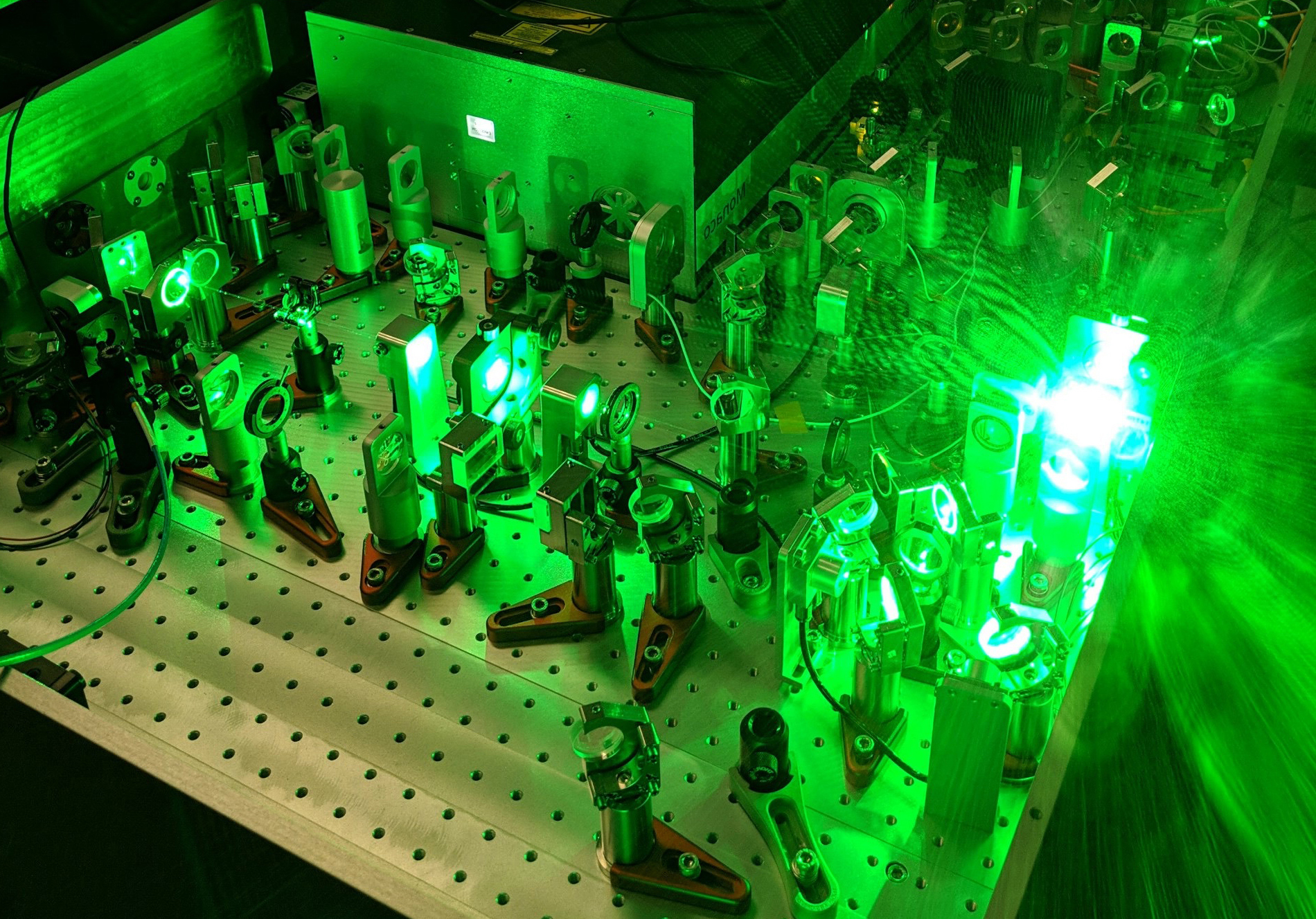
Optical layout and laser light propagation used for three-photon microscopy.
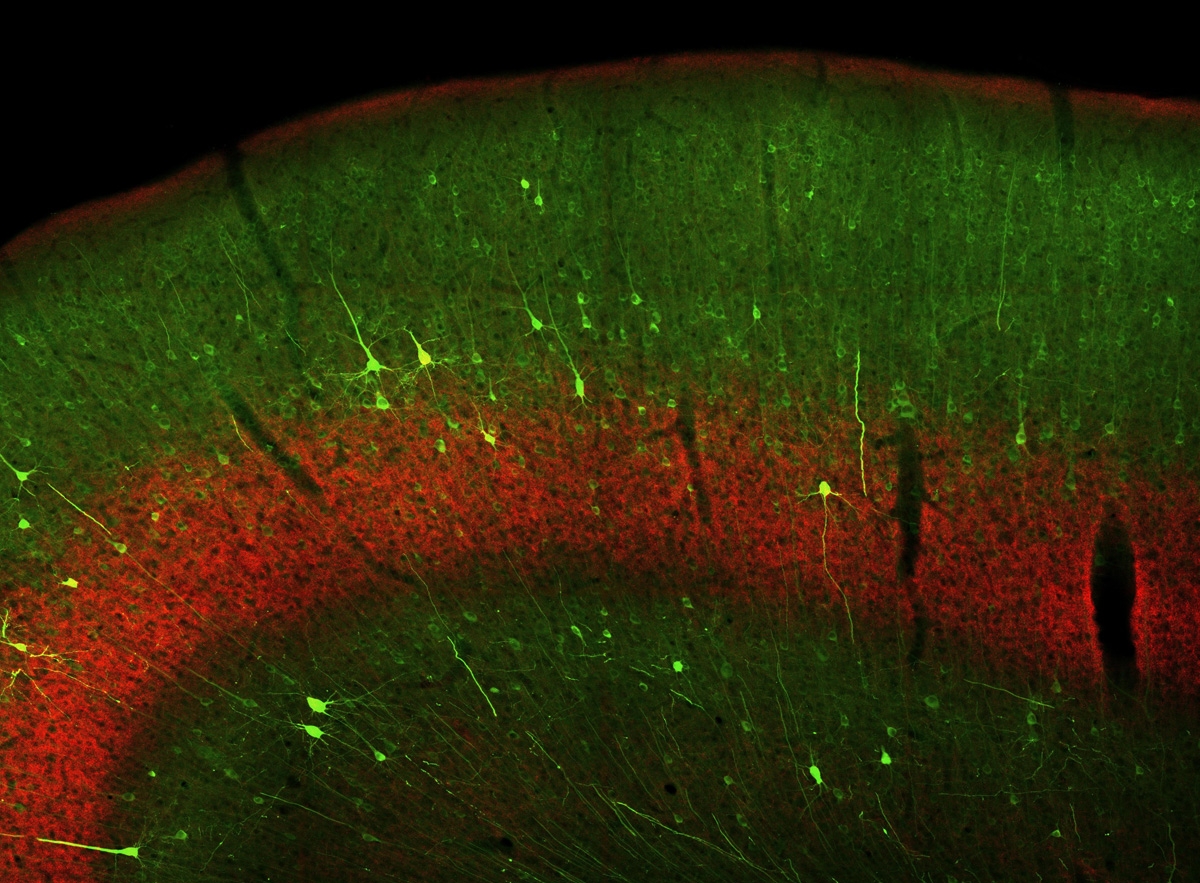
Two-photon image of the neocortex.
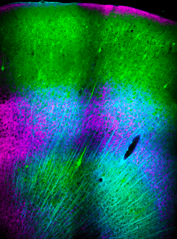
Three-photon image of the visual cortex.