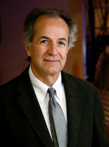
Our Team

Kamil Ugurbil , PhD
Director of the Optical Imaging MDT
Professor of Radiology, Medicine (Joint Appointment), and Neuroscience (Joint Appointment)
Director, Center for Magnetic Resonance Research
Faculty, PhD Program in Medical Physics
Faculty, Graduate Program in Neuroscience


Bio
Kamil Ugurbil currently holds the McKnight Presidential Endowed Chair Professorship and is the founding Director of the Center for Magnetic Resonance Research (CMRR) at the University of Minnesota. After completing his B.A. and Ph.D. degrees in physics, and chemical physics, respectively, at Columbia University, New York, N.Y., Prof. Ugurbil joined AT&T Bell Laboratories in 1977, and subsequently returned to Columbia as a faculty member in 1979. He was recruited to the University of Minnesota in 1982 where his research in magnetic resonance led to the evolution of his laboratory into an interdepartmental and interdisciplinary research center, the CMRR. His primary research focus has been the development and application of MR methods and instrumentation towards obtaining high spatiotemporal resolution and high accuracy functional and anatomical information in the human brain, and the development of ultrahigh magnetic fields for human imaging for biomedical research in general. This body of work has culminated in pioneering accomplishments, such as the co-introduction of functional brain imaging (fMRI), the introduction and development of ultrahigh magnetic fields (defined as ≥7 Tesla), functional mapping of columnar and layer specific functional responses in the human brain, highly accelerated functional brain imaging, and MR spectroscopy for studies of metabolism in vivo. He was one of the two PI’s of the Human Connectome Project and one of the fourteen members of the first BRAIN Initiative working group. He was recognized by several awards and honors including membership in the US National Academy of Medicine, American Academy of Arts and Sciences, Richard R. Ernst Gold Medal, ISMRM Gold Medal, ISMAR Prize, Koç Award, the IEEE Medal for Innovations in Healthcare Technology, and two honorary doctorates.
Research Summary
Kamil Ugurbil's central research interest is tackling biological problems, particularly in the brain, with new and transformative imaging technologies that involve instrumentation, image acquisition and reconstruction methods. His research is characterized by development of new technologies, and applications of these technologies, to obtain new and previously unavailable information about biological processes. This central interest was initially focused on developing, for the first time, new magnetic resonance (MR) spectroscopy methods to monitor intracellular chemistry in intact biological systems, using systems such as bacteria in suspension and perfused organs. This work pioneered the general field of using MR for the study of biological processes in vivo. In the past three decades, his focus has predominantly been the development of ultrahigh field MR methods for human neuroimaging, particularly for imaging brain activity (functional imaging (fMRI)) and connectivity and combining these methodological and instrumentation developments with neuroscience applications in the human and animal brain to advance our understanding of brain function in health and disease.
Dr. Ugurbil's research brings together physics and instrumentation with physiology, neuroscience and neurochemistry to assess cerebral function. fMRI was first achieved simultaneously by two independent teams; one was the team he lead at the Center for Magnetic Resonance Research (CMRR) at the University of Minnesota. This development has been followed by a large body of seminal work from his laboratory on the mechanisms of coupling between magnetic resonance detected signals and neuronal activity, and development of new instrumentation and techniques to exploit this information, leading to the most advanced neuroimaging studies we have today.
The effort of his group to develop new technologies to advance neuroimaging pioneered the use of ultrahigh field (≥7 Tesla) imaging in humans, particularly (but not only) for pushing the boundaries of mapping brain function and connectivity. 7 Tesla and associated methods developed to overcome the significant challenges faced with imaging the human body at such high magnetic fields currently represent the most advanced platform used for human brain research and are now increasingly used world-wide. This effort also led to the development of instrumentation capable of human imaging above 10 Tesla for the first time (see the article The world’s strongest MRI machines are pushing human imaging to new limits).
Recently, these advances have been extended to mapping the macro-connectome of the human brain under the auspices of the Human Brain Connectome project launched by the NIH Neuroscience Blueprint initiative and continued through Human Connectome Project Lifespan project.
Education
Professional Memberships
Contact
Address
2021 6th Street SE, Minneapolis, MN 55455

Bio
Prakash Kara received his PhD from the University of Alabama at Birmingham and postdoctoral training at Harvard Medical School. His previous faculty positions were at the Medical University of South Carolina, as Assistant Professor and then Associate Professor. He came to the University of Minnesota as a Full Professor in 2018 as the first hire in a new initiative, the Medical Discovery Team (MDT) in Optical Imaging and Brain Science.
Research Summary
The Kara lab solves puzzles in sensory perception and neurovascular coupling in the mammalian brain. Our long-term goal is to obtain a comprehensive view of how the brain adapts to change in adulthood, development, and disease. The short-term goals of our current projects are to (1) resolve aspects of neural coding of binocular signals in the visual cortex, (2) build microcircuits for neurovascular coupling and (3) determine the underpinnings of functional magnetic resonance imaging (fMRI).We use two-photon and three-photon imaging, optogenetics, and electrophysiological techniques. More specifically, we assay synaptic and spiking activity along with the responses of individual blood vessels to sensory stimuli and optogenetic activation.
Contact
Address
1-166D CMRRMinneapolis, MN 55455-3007


Bio
Gordon received his B.S. from Duke University and his Ph.D. from MIT, where he worked with Dr. Mark Bear to settle a long-running debate on the mechanisms of ocular dominance plasticity by demonstrating in vivo a requirement for homosynaptic LTD in the loss of visual responses. As a post-doc with Dr. David Fitzpatrick at the Max Planck Florida Institute for Neuroscience, he developed cutting edge in vivo imaging techniques that permit following the plasticity of both single neurons and large populations across development. By applying these tools to address the seemingly contradictory effects of correlated neural activity—which both drives circuit formation but also limits sensory information—he demonstrated for the first time a developmental decrease in correlated activity within a neural population, and showed that this leads to an increase in stimulus discrimination within the population.
Research Summary
Development of visual cortical circuits How are the neural circuits that process sensory information built during the course of development? As the development of a circuit inevitably constrains its future function, addressing this question is critical to understanding both the function of mature circuits and how neurodevelopmental disorders give rise to sensory deficits. The Smith Lab uses advanced optical imaging techniques in the developing visual cortex to investigate how large populations of neurons form the networks required to process visual information. Ongoing projects include: Distributed functional networks in early development Ongoing work has shown that large-scale distributed functional networks spanning millimeters exist in visual cortex well before they can be visually driven. We're working to determine the circuit mechanisms that give rise to these early networks and guide their refinement during development. Intracortical inhibition and network formation Network function critically depends on the structure and organization of inhibition within the network, but little is known about the organization of inhibition in developing cortical networks. Using novel viral tools, we're measuring and manipulating inhibitory neurons in the early cortex. How does early SA sculpt future perception? Correlated activity in early development is a critical driver of circuit formation and future perceptual processing. We're using cutting edge optical approaches to explore the causal role of early patterned network activity in visual processing, and determine whether abnormal spontaneous activity is a common theme linking neurodevelopmental disorders.
Contact
Address
2021 6th St SEMinneapolis, MN 55455-3007


Research Summary
Our lab studies the computations that make it possible to see and think. Taking inspiration from the tools and concepts of AI, we use computational models to bridge observations of brain activity at many spatial and temporal scales, from fMRI in humans to 2-photon microscopy in animal models.We are especially interested in the generative capabilities of the visual system. Much of life is spent imagining or dreaming of internal images that one has never actually observed. Why is the visual system so good at generating images, and how does this remarkable ability help us to see? We are addressing this question by monitoring the human brain as it engages complex, real-world scenery and as it calls upon memory to generate mental images.


Research Summary
As we learn a new skill, how does our brain change to store information about the actions involved? Cortical neurons receive information from the thousands of synaptic inputs onto the dendritic tree. Learning occurs when the efficacy of these inputs change in response to patterns of activity. Within the dendrite, location-dependent learning rules and interactions between inputs sculpt these changes.The Kerlin Lab uses advanced two-photon microscopy techniques to understand how dendritic compartments and individual synapses within the motor cortex are modified as mice learn to perform new tasks. Recent work has identified a distinct loop between cortex and thalamus that maintains motor plans in the absence of overt action. By tracking and manipulating dendritic activity while monitoring the kinematics of action, we are determining the critical subcellular loci for the learning of new motor plans. Clarifying the biophysical events that drive normal plasticity will help us identify ways to shift cortical plasticity into regimes that favor the improvement of cognitive motor disorders or rehabilitation after damage to motor systems.
Contact
Address
3-114 Jackson HallMinneapolis, MN 55455-0263


Bio
Madhu Kannan is an Assistant Professor of Neuroscience and a member of the Medical Discovery Team on Optical Imaging and Brain Science.
Dr. Kannan completed her undergraduate studies in Biochemistry in Chennai, India, and acquired a Master’s in Molecular Genetics at the University of Leicester, UK. She then obtained a Ph.D. in Neurophysiology at the Max Planck Institute of Experimental Medicine and George August University, in Germany. During her postdoctoral work, in the lab of Dr. Mike Higley at Yale School of Medicine, CT, she studied the mechanisms of experience-dependent plasticity at cortical inhibitory synapses using slice electrophysiology and optogenetics. She subsequently joined the lab of Dr. Vincent Pieribone, for a second postdoc, where she created a suite of mutually compatible recombinant voltage indicators and used them to perform some of the first recordings, with millisecond resolution, of the correlated dynamics of genetically distinct neuron types in cortical microcircuits in behaving rodents.
1. C Huang†, J Luo, S J Woo, L A Roitman, J Li, V A Pieribone, M Kannan†, G Vasan†, M J Schnitzer†. Dopamine signals integrate innate and learnt valences to regulate memory dynamics. Research-Square; under revision in Nature. †Corresponding authors.
2. M Kannan*†, G Vasan*†, S Haziza*, C Huang, R Chrapkiewicz, J Luo, J A Cardin, M J Schnitzer† and V A Pieribone†. Dual-polarity voltage imaging of the concurrent dynamics of multiple neuron types. Science, 378(6619), 2022. *Equal contribution. †Corresponding authors.
3. M Kannan, G Vasan, and V A Pieribone. Optimizing strategies for developing genetically encoded voltage indicators. Frontiers in Cellular Neuroscience, 13:53, 2019.
4. M Kannan*, G Vasan*, C Huang, S Haziza, J Z Li, H Inan, M J Schnitzer, and V A Pieribone. Fast, in vivo voltage imaging using a red fluorescent indicator. Nature Methods, 15(12):1108–1116, 2018. *Equal contribution. Highlighted in Yale News.
5. M Kannan, G G Gross, D B Arnold, and M J Higley. Visual deprivation during the critical period enhances layer 2/3 GABAergic inhibition in mouse V1. Journal of Neuroscience, 36(22):5914–5919, 2016.
6. A Matz, S Lee, N S Domeyer, D Zanini, A Holubowska, M Kannan, M Farnworth, O Jahn, M C Gopfert, and J Stegmüller. Regulation of neuronal survival and morphology by the E3 ubiquitin ligase RNF157. Cell Death and Differentiation, 22(4):626–642, 2015.
7. C Mukherjee, A Holubowska, N S Domeyer, M Mitkovski, S Lee, M Kannan, A Matz, M Vadhvani, and J Stegmüller. Loss of the neuron-specific f-box protein FBXO41 models an ataxia-like phenotype in mice with neuronal migration defects and degeneration in the cerebellum. Journal of Neuroscience, 35(23):8701–8717, 2015.
8. M Kannan, S Lee, N S Domeyer, and J Stegmüller. The E3 ligase Cdh1-anaphase promoting complex operates upstream of the E3 ligase Smurf1 in the control of axon growth. Development, 139(19):3600– 3612, 2012.
9. M Kannan, S Lee, N S Domeyer, T Nakazawa, and J Stegmüller. p250GAP is a novel player in the Cdh1-APC/Smurf1 pathway of axon growth regulation. PLoS One, 7(11), 2012.
10. M Kannan, J R Steinert, I D Forsythe, A G Smith, and T Chernova. Mevastatin accelerates loss of synaptic proteins and neurite degeneration in aging cortical neurons in a heme independent manner. Neurobiology of Aging, 31(9):1543–1553, 2010.
Research Summary
Animal cognition is a complex dynamic process that is tightly controlled by feed-forward and feed-back mechanisms and involves the concerted action of multiple distinct excitatory and inhibitory neuron types in the brain. Modern genetic and optical tools enable the targeted identification, stimulation, or recording of a single neuron type at a time. However, how the activation dynamics of multiple neuron types converge in real-time and how these time-varying interactions impact network output to influence the cognitive outcome are unknown. Using single-neuron, single-spike resolution, multi-population voltage imaging in mice, during selective attention paradigms adapted from primates, my research program will examine the synergistic dynamics of targeted cortical neurons in behavior and cognitive function. We will further combine this approach with targeted gene perturbation to understand the contribution of risk genes to impaired circuit function, which may in turn contribute to some of the cognitive deficits associated with psychiatric illnesses.


Creative Activity Summary
Neuronal activity underlying behavior is encoded by distinct populations comprising a myriad of excitatory and inhibitory cell classes. These populations not only form local networks in individual brain areas but also act concertedly with neural assemblies in other relevant brain areas via global connections that facilitate interareal communication. In my recent work, I developed a high-throughput protein engineering and voltage screening approach to create a suite of 4 mutually compatible fluorescent voltage indicators for recording neuronal activity at submillisecond precision. Using a novel dual-polarity, dual-color voltage imaging technique, these indicators enable concurrent optical recordings of the spiking dynamics of three distinct excitatory and interneuronal populations in the visual cortex or the hippocampus in running mice. In a parallel study, I used genetically encoded calcium indicators and a synchronized dual microscope system to uncover the concerted spatial and temporal odor patterning in the dorsal and previously inaccessible lateral regions of the olfactory bulb. Collectively, the multipopulation and multiareal recording approaches will empower studies of the interplay between local and global neural circuit dynamics underlying stimulus-guided behavior in awake animals.