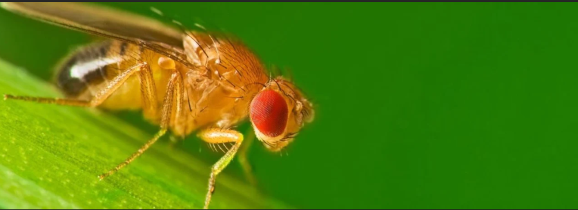
Fruit Flies Forever (but not frozen)
Drosophila melanogaster – the humble fruit fly – has long been a giant in the world of biomedical research.
According to Tom Hays, professor of genetics, cell biology and development at the U of M’s College of Biological Sciences, about 75% of human genes have a “homolog” in Drosophila. Studying those genes and how they function in Drosophila can help decipher mechanisms underlying human disease, which in turn can lead to new pharmaceuticals and other treatments.
Not surprisingly, researchers need access to fly stocks with consistent genetics. Even when fly husbandry procedures are rigorously followed, the fly lines are still subject to genetic drift via random mutations. This drift can severely complicate genetic research of disease pathways.
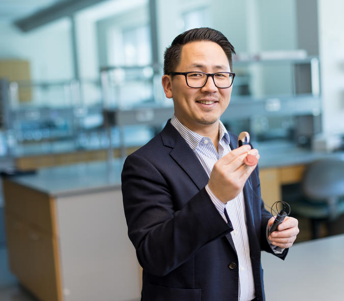
Along with Hays and other U of M researchers, IEM director John Bischof has been working on a solution. Bischof, who’s also a professor in the U of M’s departments of mechanical engineering and biomedical engineering, has been developing a method for “vitrifying” Drosophila embryos – that is, preserving them in a “glassy”, ice-free state in -196 °C (-320 °F) liquid nitrogen – and then rewarming them, again without any lethal ice formation. If the protocol can be easily adopted, it could hasten the development of new medical innovations.
Engaging “the fly community”
“I’ve been in the fly community since 1985,” says Hays, whose lab has been using Drosophila to study the genetics underlying the transport of organelles and molecular cargoes in neurons as a way of understanding neurodegenerative diseases. Keeping the “babies” happy, as Hays puts it, is “a huge labor cost.” Stocks need to be transferred to new food every two weeks, a regimen that must be maintained regardless of holidays, weekends, or other time off. “It’s not a challenging mental exercise,” Hays jokes, but it’s one “you can’t screw up.” And even when avoiding contamination during stock transfers, those lines are still subject to genetic drift.
These are problems that Bischof, who has “been intrigued with Drosophila since my PhD,” believed cryopreservation might be able to solve. Since being awarded his mechanical engineering doctorate in cryobiology at the University of California Berkeley, Bischof has developed protocols for vitrifying tissues and even whole organisms, including the first fish to develop from a vitrified embryo. Cryopreservation, he says, “can lock down the genetic information at a particular time point.” It would also be “a valuable safety precaution” for a research lab’s “absolutely fundamental lines.”
In pursuing fruit fly cryopreservation, Bischof wasn’t venturing into uncharted territory. In 1992, two scientists published two different protocols for cryopreserving Drosophila. “It made a big impression on me,” he recalls. However, “no one could repeat the protocols,” and Drosophila cryopreservation research languished.
Building on more than 25 years studying cryoprotective agents (CPAs) as a means to cool and warm biological systems without ice formation, Bischof turned to Drosophila as the National Institutes of Health (NIH) and the “fly community” in general began to push for some kind of reliable preservation technology. Not being a biologist, he needed a researcher with deep expertise in fruit fly biology and an established “fly lab” for experimental specimens. In 2019, he approached Hays, and the following year, they received a NIH grant to pursue work on Drosophila embryo cryopreservation.
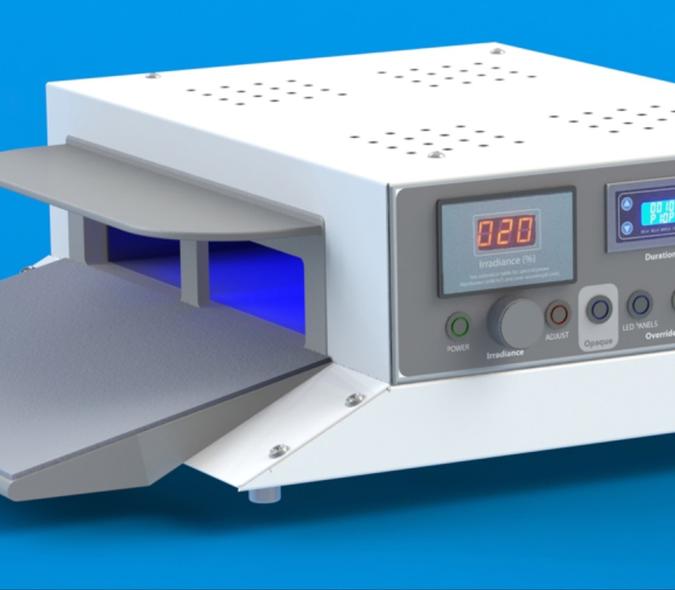
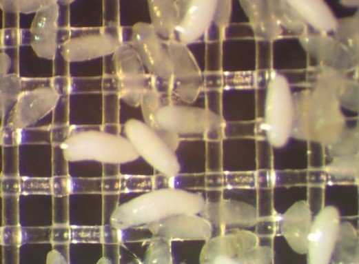
Bischof credits former U of M graduate student Li Zhan for “finding out where the failures are” in the 1992 protocols and “coming up with some clever innovations to make them work better.” Very simply, Zhan used a mesh system for storing the embryos while perfecting the timing of CPA exposure. He and Hays were then able to develop what Bischof describes as a “robust, repeatable protocol” on 25 different fly lines. “We were among the first to show not only new ways to rewarm, but also precisely what CPA concentrations were needed to make sure this was successful,” Bischof says.
The next generation
Last year, Bischof and his research colleagues were awarded a second, much larger NIH grant to further develop and share the protocol. Indiana University’s Bloomington Drosophila Stock Center, which maintains more than 86,000 unique Drosophila stocks for researchers worldwide, is generating strains that the U of M cryopreservation team will use in their experiments.
Kevin Cook, a senior scientist in Indiana’s Department of Biology and the lead principal investigator at the Bloomington center, is intrigued though somewhat skeptical. From his point of view, the current method of maintaining fly stocks is less expensive than cryopreservation, which “would require new equipment and materials.” He also believes that cryopreservation needs to prove that it can prevent or at least minimize genetic drift.
Still, Cook does see potential applications for the maintenance and backup of valuable Drosophila lines. “There’s the possibility that it could revolutionize the way that labs work,” he says. “We’ll just have to see.”
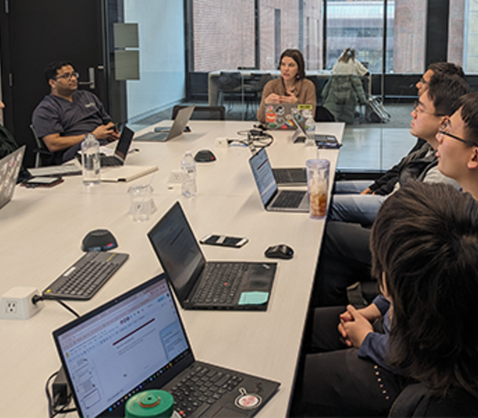
Since receiving the second NIH grant, the U of M team has been testing different meshes, CPAs, and reagents to make the protocol more robust and easily adoptable. According to Amanda Neisch, a research assistant professor who is leading the new project under the supervision of Hays and Bischof, the team will also analyze whether mutational changes occur in Drosophila after cryopreservation. “This will be the foundation for the next three years, when we will help Bloomington and other stock centers set up cryopreservation equipment and storage facilities for their Drosophila lines,” says Neisch.
Bischof acknowledges that “we’re not yet sure what the commercial aspects of this are.” (One possibility: creating cryopreservation “kits” for labs.) One thing he is certain of: “This is a great example of a team science project.” Working across their respective disciplines, biologists and engineers are addressing big challenges involving one of the smallest but most crucial biomedical research “partners.”