International Collaboration on Patellofemoral Instrumented Stress-Testing Device Provides Accurate, Objective Information
Collaboration between physicians and scientists is an essential element of advancement and innovation in orthopedics. Elizabeth Arendt, MD, professor and department vice chair, is a world-renowned orthopedic surgeon and patellofemoral expert whose research has generated international interest.
Ana Leal, PhD, is a biomedical engineer, innovation strategy lead for medical devices, and postdoc research fellow in the cMEMS group (Centre for Micro-Electro-Mechanical Systems) at the University of Minho in Portugal. Her interest in Arendt’s work led her to study at the University of Minnesota on a three-month grant. Leal and her senior clinical advisor João Espregueira-Mendes, MD, PhD, have been working over the last four years to create and validate a new device for patellofemoral (PF) instrumented stress-testing.
“The great resources offered by the University of Minnesota, from staff, research tools, and equipment (namely CMRR), bounded with Dr. Arendt’s team readiness, know-how, and experience in this specific field helped to materialize and focus this study on what should be done (patient selection, methodology definition, results interpretation), which had a very positive ‘leading’ impact in the whole project,” Leal says.
Currently, PF instability is partially determined based upon a physical examination conducted by an orthopedic surgeon and dictates what surgical route will be pursued. “The challenge is that there’s quite a bit of variability between individual exams and how we evaluate and report that, particularly in the literature, to other physicians,” explains Marc Tompkins, MD, associate professor of orthopedic surgery and co-author. “The idea is: is there any way that we could use some kind of instrument during examination testing and have more precision identifying some of the factors that we’re looking at?”
This question and the Porto Patellofemoral Testing Device (PPTD) are validated in the paper “A new device for patellofemoral instrumented stress-testing provides good reliability and validity,” published in Knee Surgery, Sports Traumatology, Arthroscopy, with Arendt, Tompkins, and the team of researchers in Portugal as co-authors.
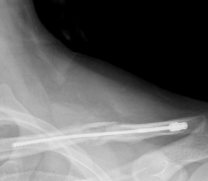
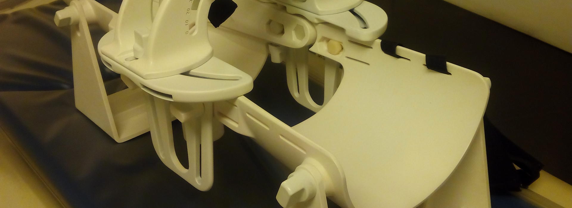
How does the device work? The PPTD holds the patient’s leg in place and applies mechanically activated force using an air pump system which moves the patella in multiple directions at various amounts of force. Since it’s made out of polyurethane, it can be used inside an MRI or CT scan, allowing the surgical team to better evaluate PF instability.
During a simple physical examination, a physician will hold the patient’s leg straight and move the kneecap from side to side, rating the movement based on the four quadrants of the patella. If there is excessive patellar movement given the amount of force applied, the doctor may determine that the medial stabilizers may not be working as well.
“The movement is highly variable in terms of how it gets interpreted,” Tompkins explained. “Some physicians are a little softer with the movement and some are more aggressive.”
The PPTD does the same thing in the MRI, however the amount of force applied and a corresponding radiographic image mitigate the subjective nature of a physical exam.
“The positive part is that it gives you the possibility of objectively measuring the patella in six planes,” Arendt says. “The negative part is that it’s used inside an MRI and so there’s a cost. For us, at least, it took a technician to do it.”
To validate the PPTD, six random patients from the University of Minnesota Medical Center with unilateral PF instability were evaluated manually by Arendt and a junior physician who were blinded to each other’s examination. The patient’s contralateral uninjured side was used as the control. The same examination was then performed using the PPTD. To examine reliability, eight volunteers without PF instability were examined under the same circumstances at the Clínica do Dragão, Espregueira-Mendes Sports Centre-FIFA Medical Centre of Excellence in Portugal.
The paper provides validation for the PPTD and shows that the device is accurate in terms of providing objective and precise data that is consistent from patient to patient.
“We are in the process of submitting another paper where we looked at different patellofemoral patient populations: patients with patellofemoral pain, patients with no instability but radiographic risk factors that would put them at risk for patellar instability, and then patients with documented patellar instability,” Tompkins added. “As you might expect, we found differences in those patient populations. But bottom line, the device is sensitive to pick that up. With that information, it is reasonable to use the device clinically.”
Arendt explained that for the device to be used clinically on a wider scale, a protocol could be developed so that physical therapists and radiology technicians could set up the device and use it correctly to evaluate patients.
“It still requires some coordination to get the patient to an MRI with the device, have somebody there to use the device and apply it appropriately, and then get the measurements and do the MRI in the correct way,” Tompkins says.
Although broad application of the PPTD is still on the horizon, collaboration between the University of Minnesota Department of Orthopedic Surgery and the researchers in Portugal will surely impact how patients with PF instability are evaluated in the future.
“I think one of the coolest parts about this was the international collaboration, as well as the collaboration between engineering and clinical orthopedics,” Tompkins says. “When people with different skills come together, some really productive work can be accomplished.”
Leal continues to collaborate with Arendt’s team around this project and related publications.
“I'd be honored to further collaborate with the University of Minnesota and contribute to medical device innovation,” she says. “There are many unmet patient needs and challenges, so the continuous cooperation between clinical staff and biomedical engineers is a must, and a personal passion.”