Medical School Research Team is First in the World to Use 2D Long Film Imaging
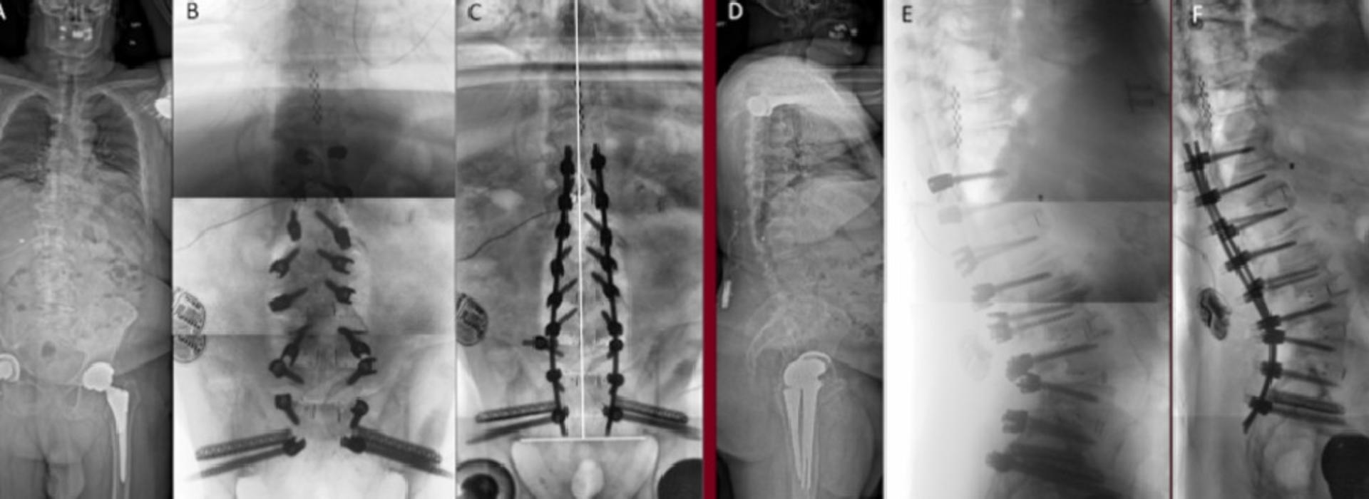
Before, during and after spine surgery, surgeons rely on clear, quality imaging to place implants in the ideal location and assess the outcome. These surgeons are also concerned with the spine’s alignment before and after the procedure, which requires a long X-ray image capturing the entire area. Typically, they use a portable imaging machine called an O-arm, however they can’t capture the entire spine in one image. Instead, they have to piece together multiple spot images to get an idea of what the whole spine looks like, which comes with workflow and image quality challenges.
The World Neurosurgery article, “2D Long Film O-arm imaging, an alternative when intraoperative fluoroscopy is inadequate,” co-authored by fifth-year resident Bryan Ladd, MD, and Assistant Professor Kristen Jones, MD, both in the Department of Neurosurgery, and David Polly Jr., MD, professor in the Department of Orthopedic Surgery, explores a new functionality for Medtronic’s O-arm that allows physicians to take clear, low-radiation long images and improves accuracy.
“Instead of stitching together multiple spot images, the tool can take a continuous X-ray, which has given us better intraoperative imaging to assess the accuracy of what we’ve done,” Dr. Polly said.
Their paper found that the new technology offers better quality and clarity, in addition to the novel ability to capture long X-ray images. Research from Medtronic also found that it reduces exposure to radiation. Dr. Polly and his team have been collaborating with Medtronic in the discussion, development and testing of the O-arm from its inception.
“Because of that partnership, we were the first in the world to use the technology that’s a part of the O-arm,” Dr. Polly said.