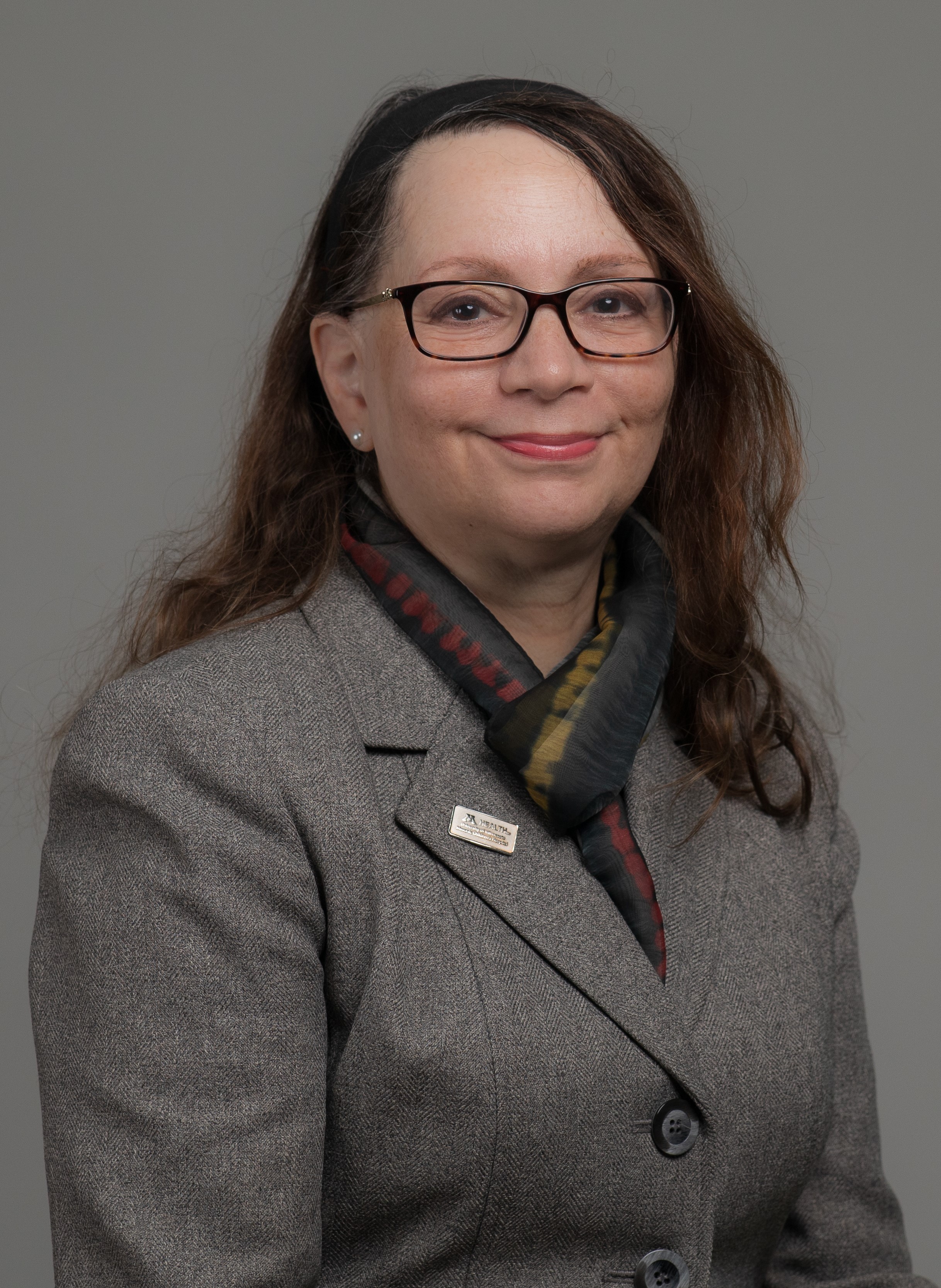
Stem Cell Institute Leadership Team

Research Summary
My complete research portfolio is now extremely collaborative using the strength of team science to drive success of investigations designed to find new approaches to disease modeling and treatment. I am the PI of a project using miPSC-derived limb bud progenitors to improve limb and digit regeneration and the Team PI of an NEI-funded RO1 with Prof. Ferrington in the Department of Ophthalmology generating an extensive biobank of iPSC-derived RPE from patients and eye donors with Dry Age-related Macular Degeneration. These iPSC-RPE provide an excellent experimental model to study mitochondrial dysfunction in AMD and for screening compounds that may maintain or restore mitochondrial function in the RPE of patients with AMD. I have also developed novel accelerated neural induction protocols to efficiently generate hiPSC-derived neuronal and glial cells for testing regenerative strategies to treat neural injury and disease. The experience from this work is directly relevant to the current Harrington proposal where we will utilize a similar protocol to generate the cortical neurons from multiple hiPSC lines for drug screening. The research team I lead also works on a range of other team projects including using iPSCs and blastocyst complementation to generate liver cells, using iPSCs and automation to investigate DMSO-free cryopreservation strategies for stem cells and stem cell-derived products and we are working with the Lions Gift of Sight Eye bank in Minneapolis to derive limbal stem cells from hiPSCs to support potential clinical use of these cells to repair corneal damage.
Contact
Address
2-226 MTRF2001 6th St SE
Minneapolis, MN 55455
Bio
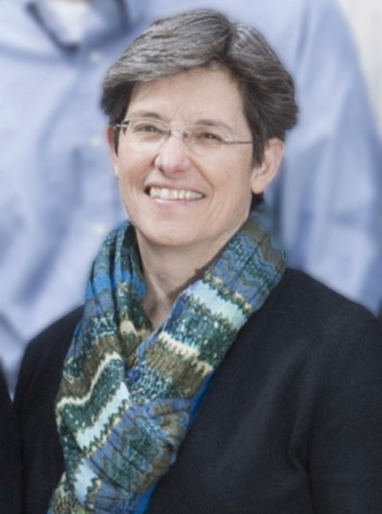
Dr. Keirstead received her Ph.D. in neurophysiology from Queen's University, Kingston, Canada, where she studied the role of neck muscle motoneurons and sensory afferents in the control of head movement in the laboratory of Dr. P. Ken Rose. As a post-doctoral fellow in the laboratories of Dr. M. Rasminsky and Dr. A.J. Aguayo at McGill University, Dr. Keirstead examined the capabilities of retinal neurons to regenerate axons and form functional synaptic connections with central nervous system neurons. Dr. Keirstead came to the University of Minnesota as a research associate in the Department of Physiology where she used calcium imaging techniques to study the regulation of intracellular calcium ion concentration in glial cells by neurotransmitters in the laboratory of Dr. Robert Miller. She continued these studies as an assistant professor in the Department of Ophthalmology.
Dr. Keirstead is an assistant professor in the Department of Integrative Biology and Physiology, and the Administrative Co-director and member of the Stem Cell Institute.
Research Summary
Current research interests include:
The functional characterization of stem cells at various stages of differentiation and their functional integration into host tissue after transplantation.
My research involves the use of calcium imaging and electrophysiological techniques to examine the functional characteristics of stem cells in vitro as they differentiate into cells of various tissue types. This system provides a useful model for the development of functional characteristics of neurons and other cells in culture. Furthermore, our functional studies will permit us to better define the optimum degree of differentiation for the successful integration of transplanted stem cells into target tissues of host animals.
Education
Contact
Address
2-2102001 SE 6th St.
Minneapolis, MN 55455

Bio
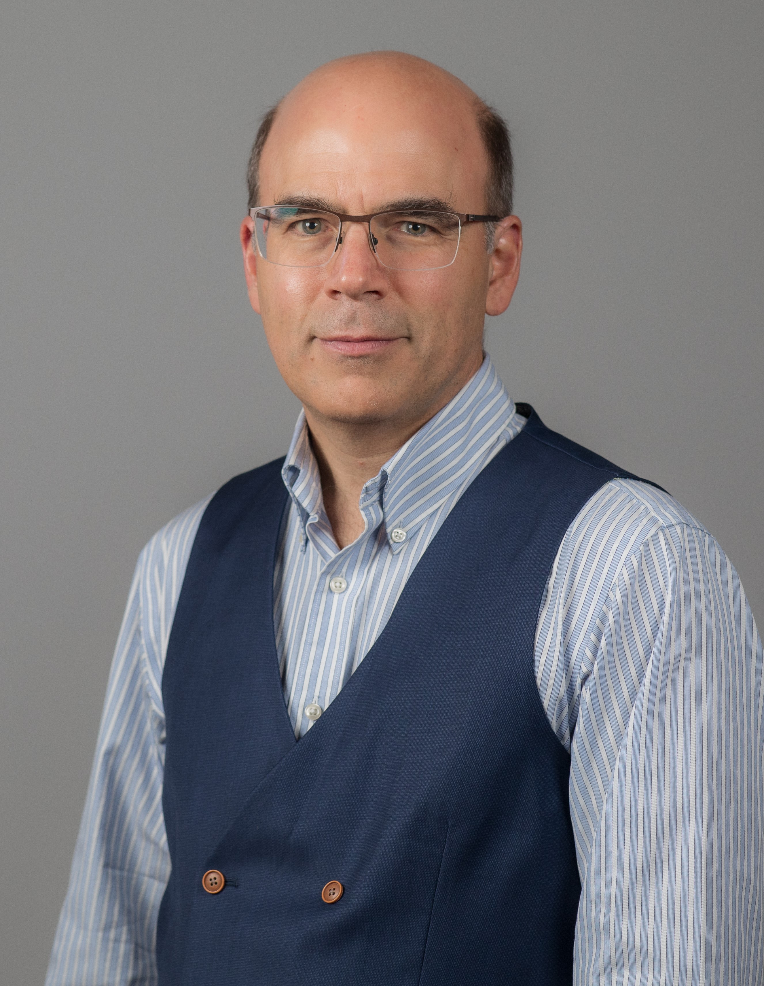
Dr. Kyba is a Professor of Pediatrics and Carrie Ramey / CCRF Endowed Professor in Pediatric Cancer Research in the Department of Pediatrics' Division of Blood and Marrow Transplant & Cellular Therapy. He is also an Endowed Scholar of the Lillehei Heart Institute, and an affiliate member of the Stem Cell Institute.
Dr. Kyba received his PhD degree from the University of British Columbia in 1998, and completed a postdoctoral fellowship in stem cell biology at the Whitehead Institute at MIT, Cambridge, MA in 2003. From 2003-2008, he was Assistant Professor of Developmental Biology at the University of Texas Southwestern Medical Center at Dallas, TX. He joined the faculty at the University of Minnesota in July 2008.
Dr. Kyba has published over 100 research manuscripts in scientific journals, including: Cell, Science, and Nature Medicine.
Research Summary
Dr. Kyba's research laboratory focuses on regulation of tissue-specific stem cells (hematopoietic and skeletal muscle) with a view towards ex-vivo expansion and therapeutic transplantation, as well as the derivation of tissue-specific stem cells from embryonic or iPS cells. He is also developing methods of performing BMT without irradiation or chemical conditioning. He has performed seminal experiments establishing the proof of principle for hematopoietic stem cell repopulation using embryonic stem cells and maintains an active program in the development of gene-targeting / genetic correction / cell therapy models.
Deriving therapeutic hematopoietic stem cells from embryonic stem cells.
ES cells are totipotent and capable of recapitulating all of the developmental events of embryogenesis. They are therefore theoretically the ideal source of cells for regenerative therapies. However, turning theory into practice is not straightforward, and very few successful models of such therapy exist. We have developed one successful model, based on regulated expression of members of the Hox family of transcription factors. Current work is focused on understanding how Hox genes regulate hematopoietic stem cell self-renewal and identifying regulatory circuits under Hox control.
Skeletal muscle stem cells and FSH muscular dystrophy
Certain degenerative diseases may be the result of ineffective self-renewal or differentiation of lineage specific stem cells. We are particularly interested in Fascioscapulohumeral Muscular Dystrophy (FSHD), a dominant dystrophy associated with a contraction of 4q subtelomeric repeats. Although the condition is almost certainly caused by derepression of a gene in the vicinity of 4q, the protein products of candidate genes in this area can not be detected overexpressed in patient muscle samples. Because muscle stem cells (satellite cells) are rare, proteins overexpressed specifically in satellite cells are unlikely to be identified in patient biopsies. We are testing the hypothesis that a Hox gene embedded within the 4q repeats, DUX4, causes FSHD when derepressed in muscle satellite cells.
Stem cell biology
Our long-term goal is to understand the pathways that control self-renewal vs differentiation of stem cells and to use this knowledge to understand degenerative diseases and to design and improve cell therapies. Our work is interdisciplinary, spanning iPS cell-based and animal models involving transplantation and tracking of somatic stem cells, vector development, CRISPR and TALEN-mediated genome editing, and cell-based screening and medicinal chemistry.
Education
Fellowships, Residencies, and Visiting Engagements
Honors and Recognition
Contact
Address
Pediatric Blood and Marrow Transplantation & Cellular TherapyMayo Mail Code 366
420 Delaware Street SE
Minneapolis, MN 55455
Administrative Contact
Elizabeth Soderberg
Administrative Phone: 612-625-8319
Administrative Email: soder348@umn.edu
Administrative Fax Number: 612-626-4074
Bio
Angela Panoskaltsis-Mortari, PhD is a Professor of Pediatrics in the Division of Blood and Marrow Transplant & Cellular Therapy. She is also a Professor of Medicine in the Division of Pulmonary, Allergy, Critical Care and Sleep. Dr. Panoskaltsis-Mortari is the Director of the Cytokine Reference Laboratory, the Director of the 3D Bioprinting Facility at the University of Minnesota and Vice Chair for Research for the Department of Pediatrics.
Dr. Panoskaltsis-Mortari received her PhD from the University of Western Ontario. She was a post-doctoral fellow in the Department of Pathology at the University of Alabama and a post-doctoral research associate in the Department of Pediatrics at the University of Minnesota. She joined the University of Minnesota faculty in 1995.
Dr. Panoskaltsis-Mortari has board certification from the American Board of Medical Laboratory Immunology. She is a member of numerous immunology, pulmonary, and hematology professional societies, and the author of over 275 articles which have appeared in such publications as Advanced Materials, Journal of Clinical Investigation, Blood, Biology of Blood and Marrow Transplantation, and American Journal of Physiology (Lung, Cell. & Mol. Physiol.).
Research Summary
With 25 years of experience in animal models of stem cell transplant, lung injury, mesenchymal stem/stromal cell therapy and the biology of graft-vs-host disease (GVHD) after bone marrow transplant, Dr. Angela Panoskaltsis-Mortari's work has evolved into the bioengineering field, and she is recognized as one of the thought leaders in lung bioengineering. Dr. Panoskaltsis-Mortari's laboratory research currently focuses upon 2 major themes: 1) bioengineering autologous tissues such as trachea and esophagus using 3D bioprinting and customized hydrogels including decellularized extracellular matrix; and 2) 3D bioprinting of cancer models.
Dr. Panoskaltsis-Mortari established and directs the 3D Bioprinting Facility at the University of Minnesota. She also directs the UMN Cytokine Reference Laboratory (a CLIA-licensed facility). She is a member of the Stem Cell Institute, the Institute for Engineering in Medicine, the Lillehei Heart Institute, the Masonic Cancer Center, the Center for Immunology, and the Robotics Institute. She is funded by the NIH, has mentored many post-docs, MD trainees, graduate students and undergrads in various training programs. Her goal is to realize the potential of regenerative medicine by converging the fields of stem cell biology, mechanical & biomedical engineering, biomaterials, physiology, robotics, and surgery to bioengineer autologous tissues/organs for transplant using a patient's own cells that would not be rejected by their immune system.
Education
Fellowships, Residencies, and Visiting Engagements
Licensures and Certifications
Honors and Recognition
Selected Publications
Contact
Address
Pediatric Blood and Marrow Transplantation & Cellular TherapyMayo Mail Code 366
420 Delaware Street SE
Minneapolis, MN 55455
Administrative Contact
Janelle Willard
Administrative Phone: 612-626-2961
Administrative Email: traut001@umn.edu
Administrative Fax Number: 612-626-4074
Bio
Our laboratory has a long-term interest in understanding the molecular mechanisms controlling lineage-specific differentiation of pluripotent stem cells (PSC), which has led to the efficient generation of PSC-derived myogenic progenitor cells endowed with in vivo regenerative potential. Current research projects focus on the effect of the environment on the engraftment and maturation of PSC-derived myogenic progenitors, the development of allogeneic and autologous cell therapy for muscular dystrophies (MD), and application of MD patient-specific iPSC-derived muscle derivatives for disease modeling and drug discovery.
Creative Activity Summary
Full list of publications at Experts@Minnesota or PubMed.
- Baik J, Ortiz-Cordero C, Magli A, Azzag K, Crist SB, Yamashita A, Kiley J, Selvaraj S, Mondragon-Gonzalez R. Perrin E, Maufort JP, Janecek JL, Lee RM, Stone LH, Rangarajan P, Ramachandran S, Graham ML, & Perlingeiro RCR. (2023). Establishment of skeletal myogenic progenitors from non-human primate induced pluripotent stem cells. Cells, 12(8), 1147; https://doi.org/10.3390/cells12081147.
- Singh BN, Yucel D, Garay BI, Tolkacheva EG, Kyba M, Perlingeiro RCR, van Berlo J, & Ogle BM, (2023) Proliferation and Maturation: Janus and the art of engineered cardiac tissue. Circulation Research, 132(4):519-540. PMCID: PMC9943541.
- McKenna DH & Perlingeiro RCR, (2023) Development of allogeneic iPS cell-based therapy: from bench to bedside. EMBO Molecular Medicine, 15(2):e15315. PMCID: PMC9906386.
- Azzag K, Bosnakovski D, Tungtur S, Salama P, Kyba M, & Perlingeiro RCR, (2022) Transplantation of pluripotent stem cell-derived myogenic progenitors counteracts disease phenotypes in a mouse model of FSHD. NPG Regenerative Medicine, 7(1):43. PMCID: PMC9440030.
- Garay BI, Givens S, Stanis N, Magli A, Yücel D, Abrahante JE, Goloviznina NA, Soliman HAN, Dhoke NR, Baik J, Kyba M, van Berlo JH, Ogle B, & Perlingeiro RCR, (2022) Inhibition of mitogen-activated protein kinase pathway enhances maturation of human iPSC-derived cardiomyocytes. Stem Cell Reports. 17(9):2005-2022. PMCID: PMC9481895.
- Kim H, & Perlingeiro RCR, (2022) Generation of human myogenic progenitors from pluripotent stem cells for in vivo regeneration. Cellular and Molecular Life Sciences. 8;79(8):406. doi: 10.1007/s00018-022-04434-8. PMCID: PMC9270264.
- Ortiz-Cordero C, Bincoletto, C, Dhoke N, Selvaraj S, Magli A, Zhou H, Kim D-H, Bang AG, & Perlingeiro RCR, (2021), Defective autophagy and increased apoptosis contribute toward the pathogenesis of FKRP-associated muscular dystrophies. Stem Cell Reports. 16(11):2752-2767. PMCID: PMC8581053.
- Dhoke N, Kim H, Selvaraj S, Oliveira NAJ, Azzag K, Tungtur S, Ortiz-Cordero C, Kiley J, Lu QL, Bang A, & Perlingeiro RCR, (2021), A universal gene correction approach for FKRP-associated dystroglycanopathies to enable autologous cell therapy. Cell Reports. 36(2):109360. PMCID: PMC8327854.
- Ortiz-Cordero C, Magli A, Dhoke N, Kuebler T, Selvaraj S, Oliveira NA, Zhou H, Sham YY, Bang AG, & Perlingeiro RCR, (2021), “NAD+ enhances ribitol and ribose rescue of α-dystroglycan functional glycosylation in human FKRP-mutant myotubes”. Elife, 2021 10:e65443. PMCID: PMC7924940
- Ortiz-Cordero C, Azzag K, & Perlingeiro RCR, (2021), “Fukutin-Related Protein: from Pathology to Treatments”. Trends in Cell Biology, 31:197-210. PMID: 33272829 (cover article).
- Kim H, Selvaraj S, Kiley J, Azzag K, Garay BI, & Perlingeiro RCR, (2021), “Genomic safe harbor expression of PAX7 for the generation of engraftable myogenic progenitors”. Stem Cell Reports, 16:10-19.PMCID: PMC7815936
- Baik J, Felices M, Yingst A, Theuer, CP, Verneris MR, Miller JS, & Perlingeiro RCR,(2020), “Therapeutic effect of TRC105 and decitabine combination in AML xenografts”. Heliyon, 6(10):e05242. PMCID: PMC7566100.
- Incitti T, Magli A, Jenkins J, Lin K, Yamamoto A and Perlingeiro RCR, (2020), “Pluripotent stem cell-derived skeletal muscle fibers preferentially express oxidative myosin heavy-chain isoforms: new implications for Duchenne Muscular Dystrophy”. Skeletal Muscle, 10(1):17. PMCID: PMC7268645
- Azzag K, Ortiz-Cordero C, Oliveira NAJ, Magli A, Selvaraj S, Tungtur S, Upchurch W, Iaizzo PA, Lu QL and Perlingeiro RCR, (2020) “Efficient Engraftment of Pluripotent Stem Cell-Derived Myogenic Progenitors in a Novel Immunodeficient Mouse Model of Limb Girdle Muscular Dystrophy 2I”. Skeletal Muscle, 10(1):10. PMCID: PMC7175515.
- Selvaraj S, Mondragon-Gonzalez R, Xu B, Magli A, Kim H, Lainé J, Kiley J, McKee H, Rinaldi F, Aho J, Tabti N, Shen W, & Perlingeiro RCR, (2019) “Screening identifies small molecules that enhance the maturation of human pluripotent stem cell-derived myotubes”. eLIFE, 8. pii: e47970. PMCID: PMC6845233.
- Selvaraj S, Dhoke N, Kiley J, Aierdi AJM, Mondragon-Gonzalez R, Killeen G, Oliveira VKP, Tungtur S, Munain AL & Perlingeiro RCR, (2019) “Gene Correction of Limb Girdle Muscular Dystrophy Type 2A Patient-Specific iPS Cells for the Development of Targeted Autologous Cell Therapy”. Molecular Therapy,27:2147-2157. PMCID: PMC6904833.
- Selvaraj S, Kyba M & Perlingeiro RCR, (2019) “Pluripotent Stem Cell-Based Therapeutics for Muscular Dystrophies” Trends in Molecular Medicine. 25:803-816. PMCID: PMC6721995. (cover article)
- Mondragon-Gonzalez R, Azzag K, Selvaraj S, Yamamoto A & Perlingeiro RCR, (2019) “Transplantation studies reveal internuclear transfer of toxic RNA in engrafted muscles of myotonic dystrophy 1 mice”. eBioMedicine. 47:553-562. PMCID: PMC6796515.
- Magli A, Baik J, Pota P, Ortiz Cordero C, Kwak IY, Garry DJ, Love PE, Dynlacht BD and Perlingeiro RCR, (2019) “Pax3 cooperates with Ldb1 to direct local chromosome architecture during myogenic lineage specification”. Nature Communications, 10:2316. PMCID: PMC6534668.
Full list of publications at Experts@Minnesota or PubMed.

Research Summary
- Molecular mechanisms of damage during freezing
- Mechanisms of cryoprotection or cryoinjury
- Low temperature imaging of biological systems
- Preservation of cells and tissues
- Microfluidics and transport in microfluidic channels

Bio
Professor and Head, Department of Biomedical Engineering
Director, Stem Cell Institute
Preceptor, Medical Scientist Training Program (Combined MD/PhD Training Program)
Faculty, Masters Program in Stem Cell Biology
Postdoctoral Fellow, Mayo Clinic College of Medicine
PhD, University of Minnesota (Biomedical Engineering), 2000
MS, University of Minnesota (Biomedical Engineering), 1998
BS, College of St. Benedict/St. John's University (Mathematics and Natural Science), 1994
Research Summary
System Regeneration Lab
The mission of our research program is to investigate the mechanisms of stem cell differentiation, especially in the context of the cardiovascular system. Driven by this mission, we also seek to generate new technologies that advance stem cell biology and promote translation of stem cell research into clinical practice. A primary strength of our program is the ability to span multiple subdisciplines within both basic science (i.e., stem cell biology, cell-cell fusion and extracellular matrices) and engineering (cytometry instrumentation and microfabrication) fields. Equally important is the high caliber of our team and the wonderful momentum they have built with hard work, strong intellect and scientific curiosity. Below is a description of the primary research projects within our research program.
I. Stem Cell Differentiation via Cell Fusion
Stem cell or progenitor cell transplantation has been proposed as a means to recover heart muscle function with damage or disease. However recent experience has taught that most transplanted cells do not successfully engraft and do not become integral components of the myocardium. Our group was among the first to discover that fusion between donor stem cells and recipient cardiomyocytes promotes stem cell engraftment, survival and differentiation. Thus, we hypothesize that cardiac differentiation of stem cells and ultimately myocardial repair after infarction might be stimulated by the transplantation of stem cells poised to fuse with cardiomyocytes of the damaged ventricle.
II. Stem Cell Differentiation via Extracellular Matrix Interactions
Extracellular matrix (ECM)-based scaffolds have been proposed as a "bio-inspired" means to deliver stem cells to damaged myocardial tissue. However, our knowledge of the impact of cell-ECM interactions on cell fate processes of stem cells is limited. Particularly lacking is a clear understanding of the impact of 3D stem cell-ECM interactions on induction or repression of differentiation. We hypothesize that ECM interactions alone are capable of driving lineage-specific differentiation of stem cells and our group is particularly interested in differentiation of cardiac cell types in this context.
III. Multiphoton Flow Cytometry to Guide Stem Cell Transplantation
The future of stem cell transplantation depends on the identification of noninvasive biomarkers to characterize cells and cell aggregates prior to transplantation. Characterization is needed minimally to define cell state and ideally to predict those cells best poised to contribute to a specific tissue type. We hypothesize that intrinsic metabolic signatures detected with multiphoton microscopy provide noninvasive biomarkers indicative of stem cell state. We have recently coupled multiphoton optics to a flow cytometry system (MPFC) that can analyze such intrinsic signatures of single cell and multicell aggregates in a high-throughput manner. This system is also the first of its kind to detect extrinsic fluorescent labels and dyes of cells within multicell aggregates or microtissues in a high-throughput manner.
