Faculty
Bio
My clinical and academic interests are focused primarily on inflammatory neuropathy. The inflammatory neuropathies include Guillian-Barre Syndrome (GBS), chronic inflammatory demyelinating polyneuropathy (CIDP), and multifocal motor neuropathy (MMN). Through various studies I hope to improve the techniques that we use to diagnose inflammatory neuropathy and to find new and improved ways to treat inflammatory neuropathy. This includes both investigation of new medications as well as improving the way we utilize standard medications. Every person is different; with different treatment requirements, different reactions to medications, and different goals for treatment. Much of my research aims to customize treatment to individual patient needs so that we can optimize treatment outcomes on an individual level.
Education
Fellowships, Residencies, and Visiting Engagements
Honors and Recognition
Contact
Address
PWB 12-100420 Delaware Street SE
MMC 295
Minneapolis, MN 55455
Administrative Contact
Clinic
Neurology Central Line: 612-626-6688
Minneapolis CSC - EMG Clinic: 612-626-6680
Academic Administrative Assistant
Cathie Witzel
witz0007@umn.edu
Bio
Dr. Atsushi Asakura is an Associate Professor of Neurology and a faculty member of the Stem Cell Institute in the University of Minnesota Medical School. He also belongs to Paul & Sheila Wellstone Muscular Dystrophy Center in the Medical School. Education: Dr. Asakura received his Ph.D. at the Institute of Medical Science at the University of Tokyo Graduate School and the National Institute of Neuroscience in Tokyo with Dr. Yo-ichi Nabeshima where he learned the molecular biology of skeletal muscle differentiation. He trained at the post-doctoral level at the Fred Hutchinson Cancer Research Center in Seattle with Dr. Stephen J. Tapscott. His post-doctoral studies involved the transcription factors for skeletal muscle development during early embryogenesis. He trained at the senior post-doctoral level at McMaster University in Hamilton and the Ottawa Health Research Institute in Ottawa with Dr. Michael A. Rudnicki where he started projects on skeletal muscle stem cells that contribute to muscle regeneration.
Research Summary
Over the past 30 years, Dr. Asakura has studied the myogenic transcription factors and muscle stem cells with an emphasis on Duchenne muscular dystrophy (DMD) therapy. Since he was a graduate student and postdoctoral fellow in the laboratories of Drs. Yoichi Nabeshima, Stephen Tapscott and Michael Rudnicki, he has focused on MyoD gene regulation and muscle stem cells more than 60 peer-reviewed publications. His laboratory's goals include attempting to understand the molecular mechanisms controlling muscle satellite cell (muscle stem cell) self-renewal and differentiation, and to develop novel therapeutic methods for DMD. This also involves the stem cell niche associated with vasculature in normal and regenerating skeletal muscle. And, he has recently begun exploration of cell-based therapy with induced Pluripotent Stem Cells (iPSCs) toward muscular dystrophy model animals.
Vascular niche for satellite cell self-renewal, muscle regeneration and muscular dystrophy therapy:
For an effective form of therapy of DMD, both the muscle and the vasculature need to be addressed. To reveal the developmental relationship between DMD and vasculature, Dr. Asakura created mdx mice, an animal model for DMD, carrying Flt1 mutation with increased vasculature (Hum Mol Genet, 2010). Using the animal models, his study is the first showing that developmentally and pharmacologically increasing vascular density can rescue the dystrophic phenotype in DMD model mice (PLoS Genet, 2019). This approach might be also effective to treat Facioscapulohumeral muscular dystrophy (FSHD) (J Clin invest, 2020. These data suggest that increasing the vasculature in DMD may ameliorate the histological and functional phenotypes associated with this disease. A patent for this technology has been granted in 2018 (US Patent, 2018). In addition, he established a tissue clearing protocol for skeletal muscle. Using this protocol, he is the first to image murine whole skeletal muscle using confocal and two-photon microscopy (Methods Mol Biol, 2016), and demonstrate that satellite cells pattern the microvasculature to be in close proximity to them via VEGFA, keeps the satellite cells in a more quiescent state via Notch pathway, suggesting a beneficial cross-talk (Cell Stem Cell, 2018).
Circadian transcriptional regulation in muscle stemcells:
Dr. Asakura has dedicated much of his professional career to studying MyoD function during myogenesis and muscle regeneration, and he has published more than 30 original research articles related to MyoD since he was a graduate student. His first contributions to science as an independent investigator were to demonstrate: 1)that MyoD negatively regulates satellite cell self-renewal (PNAS, 2007) via a MyoD-mediated induction of apoptosis through microRNA-mediated Pax3 gene suppression (J Cell Biol, 2010): 2) that the CDK inhibitor p57 plays essential roles in muscle differentiation as a MyoD downstream gene(Elife, 2018): 3) that myogenesis is regulated by an isomerase activity of Fkbp5 (Cell Rep, 2018): 4) that the core circadian regulator Cry2 and Per2 which regulates MyoD and IGF2 expression promotes myoblast proliferation and subsequent myocytefusion to form myotubes in a circadian manner (Cell Rep, 2018; bioRxiv, 2020).Therefore, his hypothesis is that genetic modification of transplanted stem cells using a modification of MyoD cascade may be utilized to effectively treat DMD and improve musclefunction.
DMD therapy via induced Pluripotent Stem Cells(iPSCs):
Dr. Asakura also demonstrates that upon being infected with a retroviral vector expressing the four reprogramming factors, the satellite cell-derived myoblasts successfully gave rise to iPSC colonies. To the best of their knowledge, this study is the first showing that fully committed myogenic cells can be reprogrammed to iPSCs (Stem Cells. 2011). His hypothesis is that myoblast-derived iPSCs may maintain epigenetic memory of myogenic status, which might contribute to the higher myogenic differentiation potential. Recently, he established the blastocyst complementation to generate iPSC-derived skeletal muscle in chimeric animals (World Patent Application, 2016). A key to the generation of human myogenic cells and skeletal muscle in a host animal is the selective knockout of genes in the blastocyst that are critical for organ development. These concerted approaches will help us to create iPSC-derived myogenic cells in vivo, which can be transplanted into patients for a definitive cure of DMD. In addition, the generation of iPSC-derived DMD skeletal muscle in mice will serve as an animal model to study the characteristics and regeneration of the human skeletal muscle diseases and responses to pharmacological agents such as exon-skipping.
Contact
Address
McGuire Translational Research Facility2001 6th St SE, Rm 4-220
Minneapolis, MN 55455


Research Summary
My work focuses on the physiology and biophysics of skeletal muscle. The work focuses primarily on probing the interactions of actin and myosin and the role of the passive resistance provided by titin protein filaments. I am interested in the relationship between macromolecular structures and their physiological functions. I the intricate organization and interaction of macromolecules in skeletal muscles and in the eye provide model systems for the study of the coupling of form and function. It is our hope that this combination of mechanical and biochemical data will help us to relate better molecular level information to the physiological responses to biological tissues.
Education
Contact
Address
3-136 Jackson Hall321 Church St SE
Minneapolis, MN 55455
Research Summary
My laboratory is primarily interested in metabolic physiology, stress physiology and social determinants of health and aging. We developed a unique integrative approach to investigate the molecular physiology of obesity and chronic stress-related disease which combines behavioral and genetic models, metabolic, neuroendocrine and imaging techniques in living animals as well as sophisticated molecular and structural biology analyses of neuropeptides.
The major research topics include: 1) the functional role of Vgf-derived peptides in obesity and metabolism and their development as innovative drug targets for obesity-related disease; 2) autonomic nervous system regulation of white and brown adipocyte functions in obesity; 3) stress-induced cardiovascular and metabolic diseases; 4) social determinants of health, disease and aging.
Funding sources include: NIDDK, NIA, NHLBI and several Foundations
Contact
Address
3-139 CCRB2231 6th St SE,
Minneapolis, MN 55455
Bio
Awards & Recognition
- Castle Connolly Top Doctors in America (2011-2022)
- Minnesota Monthly Top Doctors (2014-2022)
- Minneapolis St. Paul Top Doctors ( 2012-2022)
- Minneapolis/St. Paul Business Journal Health Care Hero (2022)
- Advocates for Better Health, Charles Bolles Bolles-Rogers Award (2021)
- Surgical Educator of the Year, University of Minnesota Department of Surgery (2018)
- Minnesota Physicians Community Caregivers Volunteer Physicians (2020)
- Defense Meritorious Service Medal (2009)
Medical School
M.D., University of Kansas, School of Medicine, Kansas City, KS
Residency
Residency, University of Kansas, School of Medicine, Kansas City, KS (General Surgery)
Fellowship
Fellowship, University of Minnesota, Minneapolis, MN (Surgical Critical Care)
Research Summary
Expertise: Surgery, Surgical Oncology, Critical Care and Trauma, Pancreatitis and Transplant
Service Summary
- Member, Clinical Trials Steering Committee, U.S. Army Institute for Surgical Research
- Reviewer, National Institutes of Health
- Vice Chair, Perioperative Services and Quality
Clinical Summary
Acute care; Critical care; High risk surgery; Islet Transplant Surgery; Minimally Invasive Surgery; Surgery (gastrointestinal, hernia, splenic, skin and soft tissue, billiary, laparoscopic, high risk, acute care, emergency, trauma surgery); Surgical infectious diseases; Surgical treatment of chronic pancreatitis; Trauma Services
Honors and Recognition
Contact
Address
MMC 195420 Delaware St. SE
Minneapolis, MN, 55455
Administrative Contact
Robyn Kim | kimx2349@umn.edu
Bio
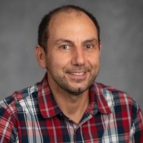
Dr. Darko Bosnakovski is an Assistant Professor in the Department of Pediatrics, Division of Blood and Marrow Transplantation & Cellular Therapy. His long-running research interest is in tissue regeneration and understanding the molecular mechanism of FSHD and sarcomas.
Dr. Bosnakovski received his DVM from Ss. Cyril and Methodius University in Skopje, N. Macedonia, and his PhD from Hokkaido University, Japan. He completed his postdoctoral training at UT Southwestern Medical Center, USA.
Education
Fellowships, Residencies, and Visiting Engagements
Honors and Recognition
Contact
Address
Office AddressLillehei Heart Institute CCRB 4-134
2231 6th St SE
Minneapolis, MN 55455
Lab Address
CCRB 4-240 BENCH 76
Administrative Contact
Sarah Jutila Peterson
Administrative Phone: 612-626-2920
Administrative Email: juti0009@umn.edu
Administrative Fax Number: 612-626-4074
Bio
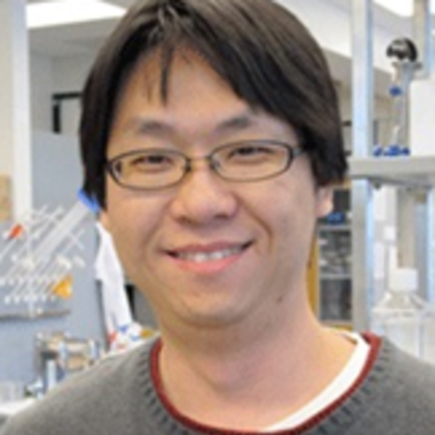
Dr. Sunny Chan is an Assistant Professor in the Department of Pediatrics, Division of Blood and Marrow Transplant & Cellular Therapy. Dr. Chan received his BSc in Pharmacology (1998) and BASc in Computer Engineering (2001) from the University of British Columbia, Canada, and PhD in Pharmacology from the Chinese University of Hong Kong in 2005. He did his postdoctoral fellowship at the National Cheng Kung University, Taiwan, and at the University of Minnesota in the laboratory of Dr. Michael Kyba. Dr. Chan joined the faculty of the University of Minnesota in 2015.
Research Summary
Dr. Chan’s research focuses on regenerative medicine for muscular dystrophies using cardiac and skeletal myogenic progenitors derived from pluripotent stem cells. He is developing methods to model cardiopharyngeal mesoderm, a cell population that forms second heart field and head muscle cells. The goal is to produce cell types appropriate for studying the physiology of muscular dystrophies specific to second heart field and head muscles in vitro and for developing cellular therapy targeting these diseases in vivo.
Education
Honors and Recognition
Contact
Address
Pediatric Blood and Marrow Transplantation & Cellular TherapyMayo Mail Code 366
420 Delaware Street SE
Minneapolis, MN 55455
Administrative Contact
Elizabeth Soderberg
Administrative Phone: 612-625-8319
Administrative Email: soder348@umn.edu
Administrative Fax Number: 612-626-4074
Bio
Administrator Info
Email: endofixer@umn.edu
Fax: 612-626-3133
Mail: 420 Delaware Street SE, MMC 101, Minneapolis, MN 55455
Summary
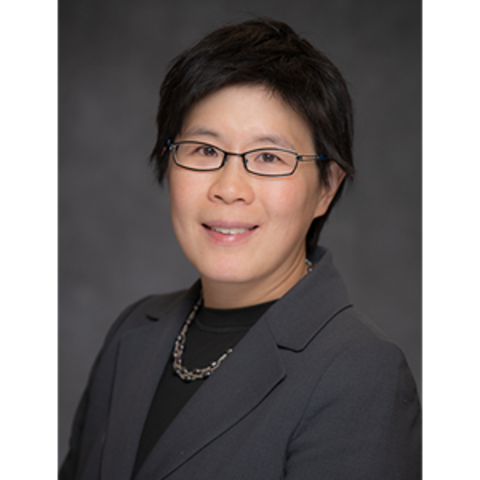
Dr. Lisa Chow finished medical school at the University of Minnesota in 1999 and completed an internal medicine residency and endocrinology fellowship at the Mayo Clinic. She joined the faculty at the University of Minnesota in 2006 and completed a Master's in Clinical Research in 2011. Dr. Chow is actively engaged in clinical research and is especially interested in the effects of lifestyle intervention (dietary changes, exercise) to treat insulin resistance and diabetes. She is also highly interested in mentoring the next generation of scientists by serving as the Director of the T32 training program for Diabetes, Endocrinology and Metabolism at the University of Minnesota.
Research Summary
The effects of lifestyle changes (diet, exercise) on insulin resistance and diabetes
Clinical Summary
Bone; Diabetes; Osteoporosis
Honors and Recognition
Selected Publications

Research Summary
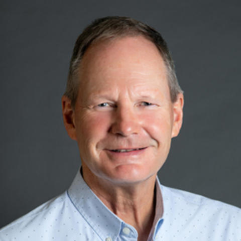
My laboratory primarily studies the structure and cellular function of the dystrophin-glycoprotein complex, which spans the muscle cell plasma membrane (or sarcolemma) and links the cortical actin cytoskeleton with the extracellular matrix. Greater understanding of the physiologic role of the dystrophin-glycoprotein complex is necessary to understand how its absence or abnormality leads to Duchenne muscular dystrophy and forms of human dilated cardiomyopathy. The lab has defined the complete actin-binding region of 400 kDa dystrophin and shown that its homologue utrophin binds actin filaments through a distinct molecular mechanism. Novel methods to visualize the sarcolemmal cytoskeleton without interference from internal structures provided the first evidence that dystrophin functions in vivo to mechanically stabilize ?-actin filaments in costameres. Studies of dystrophin-deficient mice and new animal models generated by the lab have provided insight into the function of costameres in striated muscle and suggest novel links between dystrophin deficiency and alterations in cell signaling, or gene expression manifest by dystrophic muscle. My lab's unique capability to express biochemical amounts of full-length dystrophin and utrophin has made possible new studies to i) characterize the effects of dystrophy-causing point mutations on dystrophin structure/function, ii) to identify novel associated proteins and iii) to develop new protein-based therapies for dystrophinopathies. In a completely new line of investigation, my group is working to determine the potentially unique roles of non-muscle actin isoforms in the establishment/maintenance of cell polarity in a variety of tissues. The ?- and ?-isoforms of actin distribute to distinct locations within a variety of polarized cell types, including neurons, epithelial cells, and hair cells of the inner ear yet ?- and ?-actin differ from each other by only 4 amino acids. Using new isoform-specific reagents and conditional knock-out mouse lines developed during the course of our muscular dystrophy studies, the lab is now working to identify non-overlapping functions of these two highly conserved and widely expressed proteins.
Research Summary
Myocardial Metabolism and its Relation to Cardiac Function Energy Metabolism and Ischemia
Clinical Summary
Congenital Heart Disease; Growth of Hypoplastic Ventricles; Esophageal Atresia and Tracheoeophageal Fistula

