Pushing the limits of fMRI to learn more about the brain and to help neurosurgeons improve patient outcomes
Assistant Professor Luca Vizioli, PhD, who recently joined the Neurosurgery Department, has worked extensively with functional MRI (fMRI) — both with patients and in his research. “As a cognitive neuroscientist, I’ve always been fascinated by how we can take pictures of the brain,” he said. “But fMRI goes beyond just imaging; you can see structures and you can also see the brain actively working.”
Functional magnetic resonance imaging measures brain activity by detecting changes associated with blood volume, flow, and oxygen levels. This technique relies on the fact that cerebral oxygenated blood flow and neuronal activation are coupled. When an area of the brain is in use, blood flow to that region also increases, according to Vizioli.
Jumped at the chance
When he got the opportunity in 2010 to work with high-field (3 Tesla and above) and ultra-high field (7 Tesla and above) MRI at the U’s Center for Magnetic Resonance Research (CMRR), Vizioli (pictured at left) jumped at the chance. “After coming to the CMRR, I have been working on pushing the limits of fMRI,” he said. “By limits, I’m talking about spatial and temporal resolution of the images — how we can get more precise images both in space and time.” CMRR Professor Essa Yacoub, PhD, has been pivotal in Vizioli’s scientific development, especially regarding high-field fMRI.
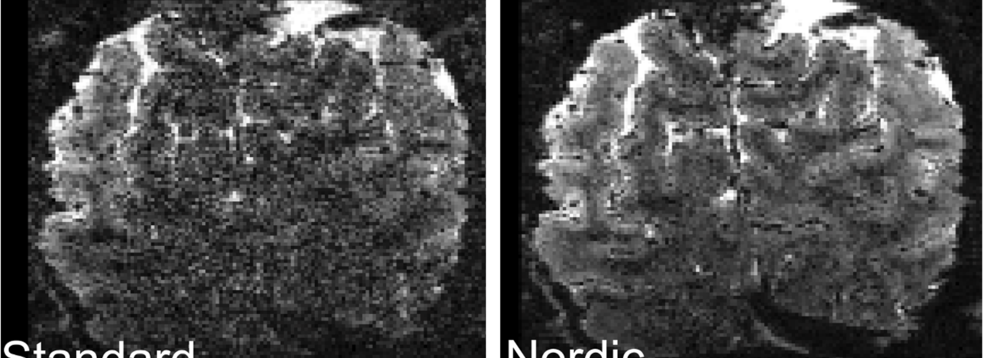
Vizioli acknowledges there are a lot of challenges in their work with fMRI. “We keep knocking down the barriers that we thought were the upper limits for these types of resolution,” he said. “As we get more precise, the images become noisier. We need to deal with the noise effectively to be able to use the images constructively.”
Noise compromises the quality and reliability of the images and represents a limit to the spatial and temporal resolution that can be achieved, according to Vizioli. “Grossly speaking there are two major types of noise sources that contaminate the fMRI signal: physiological and thermal,” he explained. “The former refers to unwanted signal originating from physiological sources, such as breathing or cardiac pulsation; the latter is caused by the MR hardware itself.”
Getting rid of the noise
There are computational algorithms that help reconstruct fMRI images to get rid of the noise, Vizioli noted. “At ultra-high field, as the resolution of functional images keeps increasing, thermal noise becomes dominant,” he said. “Some of my latest work focuses on how we can suppress thermal noise.” (See image above.) Vizioli is first author on an article1 in the journal, Nature Communications, that focuses on that topic.
He is using the fMRI experience he has gained working at CMRR under the leadership of the Center’s director, Professor Kamil Ugurbil, PhD – one of the pioneers of the field – to study the layers or laminae of the brain. “With the advent of ultra-high field MRI, we can acquire extremely precise – sub-millimeter – pictures of the brain,” said Vizioli. “This allows us to segment the cortex into its component layers and to study how each of them function.”
Impact on patients
What does all this mean for neurosurgical patients? “There are major fMRI limitations when neurosurgeons work with patients,” said Vizioli. One of the biggest factors is that a large amount of data is required to average out the noise generated by fMRI. Acquiring this amount of that data takes time. Neurosurgeons using fMRI during a procedure don’t have that kind of time. “We must produce very precise images of the brain to help direct the neurosurgeon’s interventions,” said Vizioli. “Dealing with the noise becomes very important in this situation. Getting rid of the noise in the data effectively can allows us to generate images that are just as good using less data and therefore, leads to less time that the patient has to spend in the scanner to gather that data.”
Unique in the world
Vizioli is also working with Neurosurgery Department Head Clark C. Chen, MD, PhD, to improve intraoperative MRI. “We’re one of the few centers in the world – if not the only place in the world – where we can combine all the expertise required to be successful in this endeavor,” he said. “Our preliminary data2 is really promising for bringing down the acquisition time to get the data we need.”
Vizioli believes that eventually, using the fMRI techniques being developed at the U, they will be able to build the infrastructure needed to objectively evaluate neural organization and plasticity. “We can transform fMRI into a tool that can assess tissue samples before and after surgery, evaluate brain plasticity and organization, and determine if surgery has been successful,” he said. “We have the opportunity to apply all the theoretical research we do in clinical settings.”
1Lowering the thermal noise barrier in functional brain mapping with magnetic resonance imaging