Unique national course held at the U of M teaches neurosurgery residents the demanding skills of microsurgery
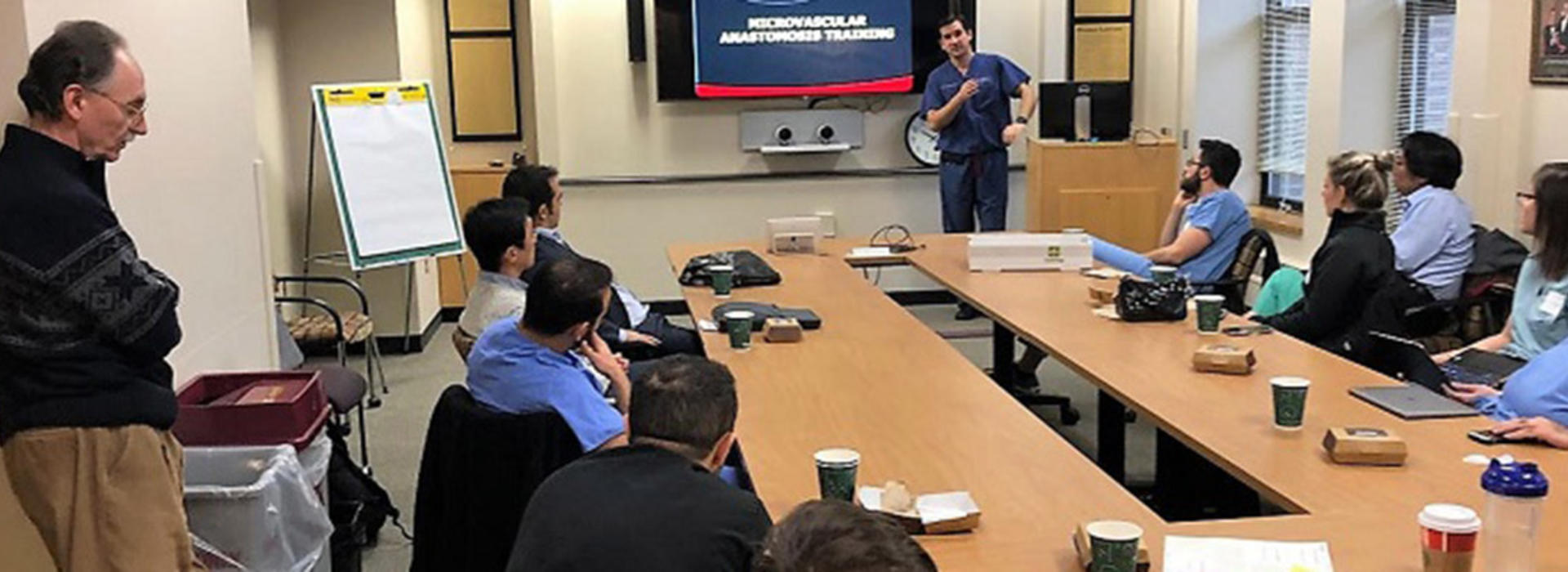
Imagine performing a surgical exercise under a microscope using sutures as thin as a human hair. That was just one of the skills taught during the first-ever microsurgery course held recently in the U of M’s Neuroanatomy Lab.
Taught by:
- Mario Zuccarello, MD, professor of Neurosurgery and director of Skull Base Surgery at the University of Cincinnati (pictured standing in the doorway)
- Andrew Grande, MD; and Ramu Tummala, MD, Department of Neurosurgery, University of Minnesota
- Ondrej Choutka, MD, FAANS, Saint Alphonsus Neuroscience Institute, Boise, ID
- Junichi Yamamoto MD, Albany Medical Center Department of Neurosurgery, NY; and
- Ulas Cikla, MD, Department of Neurological Surgery, University of Wisconsin School of Medicine and Public Health;
the course was designed to provide an almost one-to-one student/teacher ratio.
Industry sponsors of the course were: Ace Medical, Haag-Streit, Integra Lifesciences, Minntronix, Mizuho OSI, and Mutoh America Co., Ltd.
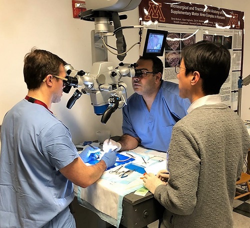
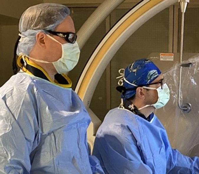
U of M neurosurgery resident, Coridon Quinn, IV, MD, (at left), performing a delicate microsurgery exercise under the watchful eye of instructor Junichi Yamamoto, MD
Attending were senior neurosurgery residents from across the country, including U of M residents, Adam Khan, MD; and Coridon Quinn, IV, MD. “This course is important because it teaches residents the basics of microsurgical skills, which is a fundamental part of neurosurgical procedures,” noted Grande. “In residency, you have daily surgical teaching, but few programs have a coordinated course within a lab in which this type of skill is taught.”
Students worked two to a station using high-powered microscopes. Most of the work was done using a small animal model, with students completing intense exercises such as detaching and reattaching tiny blood vessels or disconnecting and reconnecting a kidney.
“Another method is to use chicken wings, finding blood vessels and creating anastomosis — a cross-connection between adjacent channels, tubes, fibers, or other parts of a network,” explained Zuccarello. He noted that Cikla also demonstrated a model using the fibrous connections in a grapefruit adding, “There are many ways to develop and refine these skills.”
It’s technically demanding, according to Zuccarello, because students work on tiny structures under a powerful microscope using small precision tools. “Every movement must be well coordinated,” he noted. “And of course, you need to control your natural tremor.”
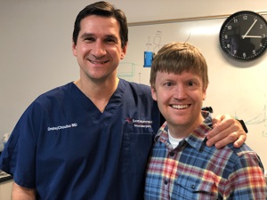
Course instructors Ondrej Choutka (at left), MD, and Andrew Grande, MD, were neurosurgical residents together at the University of Cincinnati under the tutelage of Mario Zuccarello, MD
Microsurgical skills are used every day during brain surgery as neurosurgeons work closely to important structures such as blood vessels and nerves. “These skills are fundamental for whoever works under the microscope,” Zuccarello said.
It takes training and endurance – both mental and physical – to learn these skills. “It is not something everyone can do,” Zuccarello added. “It takes a personal commitment to perfect these skills for your patients.
“Our goal was to give attendees the structure upon which they will build their surgical practice,” added Zuccarello. “You don’t become an outstanding microsurgeon just because you took a course over a day or two. You learn what you need to practice over and over in the lab and in clinical settings to constantly refine and improve these skills.”
The next Microsurgery Skills Lab is scheduled for the first week in April at the University of Cincinnati in Ohio. Many of the same instructors will be on hand, donating their time to what they see as an important addition to neurosurgery resident training.