Small Animal Imaging
Cancer and Cardiovascular Research Building (CCRB)
IVIS Spectrum In vivo Imaging System
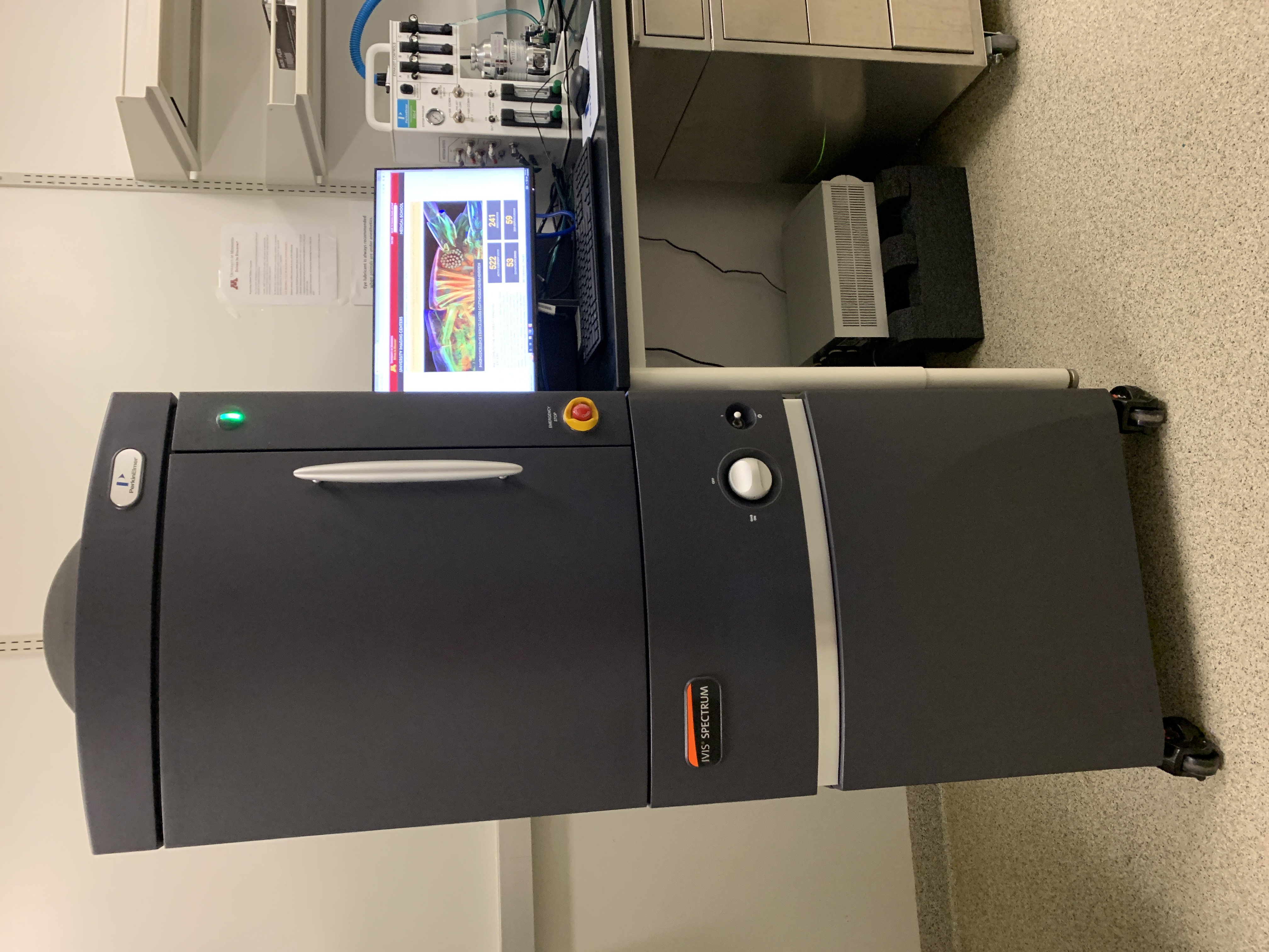
IVIS Spectrum
The IVIS Spectrum is the most versatile and advanced optical animal in vivo imaging system available on the market today. An optimized set of high-efficiency filters and spectral unmixing algorithms lets you take full advantage of bioluminescent and fluorescent reporters across the blue to near-infrared wavelength region. It also offers 3D tomography for both fluorescent and bioluminescent reporters that can be analyzed in an anatomical context using the Digital Mouse Atlas.
For advanced fluorescence imaging, the IVIS Spectrum has the capability to use either trans-illumination (from the bottom) or epi-illumination (from the top) to illuminate in vivo fluorescent sources. 3D diffuse fluorescence tomography can be performed to determine source localization and concentration using the combination of structured light and trans-illumination fluorescent images. The instrument is equipped with 10 narrow-band excitation filters (30 nm bandwidth) and 18 narrow-band emission filters (20 nm bandwidth) that assist in significantly reducing autofluorescence by the spectral scanning of filters and the use of spectral unmixing algorithms. In addition, the spectral unmixing tools allow the researcher to separate signals from multiple fluorescent reporters within the same animal.
Grant Information: The CCRB IVIS Spectrum was purchased using federal funds from NIH grant S10OD032261. All publications including data collected on the CCRB IVIS Spectrum must cite the grant.
"This work was supported by NIH grant S10OD032261 for the purchase of the IVIS Spectrum imager and by the staff at the University of Minnesota University Imaging Centers (UIC). SCR_020997."
Detector
- Andor Back-thinned, Back-illuminated CCD, cooled to -90 degrees C
- 2048 x 2048 Pixels Array, 13.5-micron sensor size
- 21.5 cm maximum image width (up to 5 mice)
Excitation
- 430-745 nm (10 filters)
- ND2 filter for trans-illumination
Emission
10 excitation filters with 30 nm bandwidth, and 18 emission filters with 20 nm bandwidth. The central filter wavelength is listed.
| Excitation | Emission |
|---|---|
| 430 | 500 |
| 465 | 520 |
| 500 | 540 |
| 535 | 560 |
| 570 | 580 |
| 605 | 600 |
| 640 | 620 |
| 675 | 640 |
| 710 | 660 |
| 745 | 680 |
| 700 | |
| 720 | |
| 740 | |
| 760 | |
| 780 | |
| 800 | |
| 820 | |
| 840 |
G8 µPET/CT
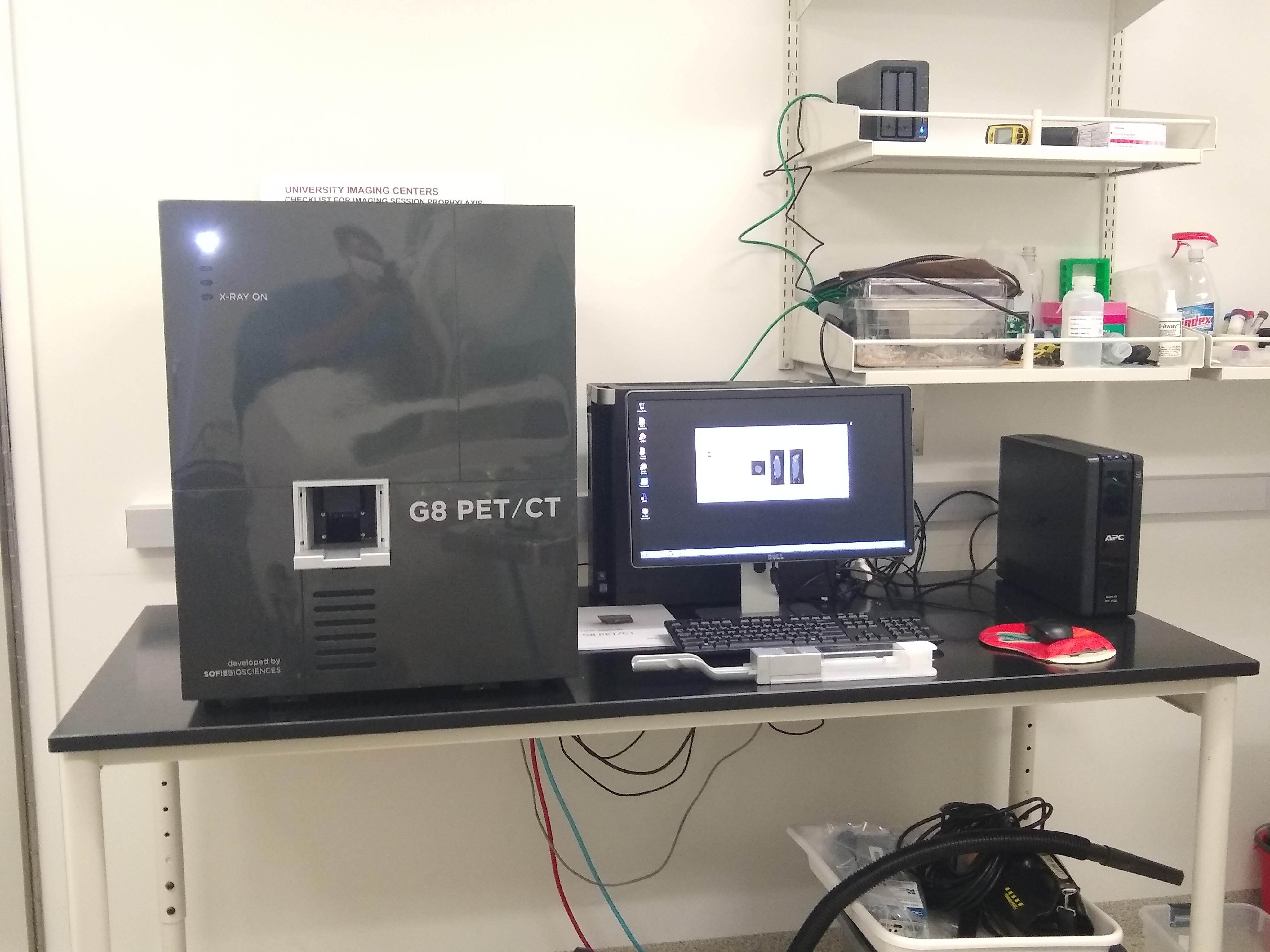
G8 PET/CT
The G8 PET/CT high-performance benchtop PET/CT imaging system offers enhanced image quality, proper animal care, safety, and increased study volume.
Employing high-resolution detectors, fast electronics, and advanced 3D image reconstruction, G8 PET / CT’s small size does not sacrifice performance (14% sensitivity, 1.4 mm resolution) or image quality.
Resolution
- 200um reconstructed CT
- 457um PET
G8 CT Specifications
| Description | Spec |
|---|---|
| X-ray Source | 59 kVp, 100uA |
| X-ray Camera | 75 micron pixels, 14bits, CMOS+Csl flat panel |
| Camera Speed | 24 fps |
| Field of View | 100mm axial by 50mm transaxial |
| Standard Scan Time | <60 seconds |
| Dimensions | 56 cm x 63 cm x 71 cm (W x D x H) |
| Weight | 90 kg |
G8 PET Performance Specifications
| Description | Spec |
|---|---|
| Axial FOV | 9.5 cm |
| Transaxial FOV | 4.7 cm |
| Detector element size | 1.8 mm x 1.8 mm x 7 mm |
| Peak absolute system sensitivity* | >14% |
| Reconstructed resolution at the center of FOV* | 1.4 mm |
| Average energy resolution | ≤18% |
| Energy window range | 150 - 650 keV |
| Total number of detector elements | 5,408 |
| Reconstruction algorithm | 3D ML-EM |
Vevo F2 Ultrasound
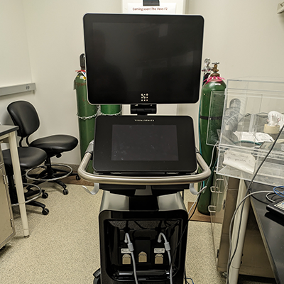
Vevo F2
Ultra-high to low frequency ultrasound (71–1 MHz) allows for imaging a range of animal models. Users now have the flexibility to image at low frequency for penetration and ultra-high frequency for resolution.
Available Licenses
Acquisition Software
Analysis Software
Jackson Hall
IVIS Spectrum In vivo Imaging System
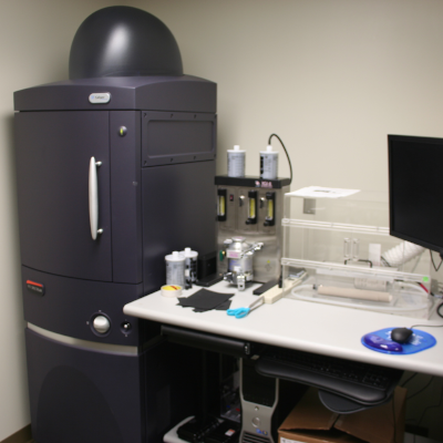
IVIS Spectrum
The IVIS Spectrum is the most versatile and advanced optical animal in vivo imaging system available on the market today. An optimized set of high-efficiency filters and spectral unmixing algorithms lets you take full advantage of bioluminescent and fluorescent reporters across the blue to near-infrared wavelength region. It also offers 3D tomography for both fluorescent and bioluminescent reporters that can be analyzed in an anatomical context using the Digital Mouse Atlas.
For advanced fluorescence imaging, the IVIS Spectrum has the capability to use either trans-illumination (from the bottom) or epi-illumination (from the top) to illuminate in vivo fluorescent sources. 3D diffuse fluorescence tomography can be performed to determine source localization and concentration using the combination of structured light and trans-illumination fluorescent images. The instrument is equipped with 10 narrow-band excitation filters (30 nm bandwidth) and 18 narrow-band emission filters (20 nm bandwidth) that assist in significantly reducing autofluorescence by the spectral scanning of filters and the use of spectral unmixing algorithms. In addition, the spectral unmixing tools allow the researcher to separate signals from multiple fluorescent reporters within the same animal.
Detector
- Andor Back-thinned, Back-illuminated CCD, cooled to -90 degrees C
- 2048 x 2048 Pixels Array, 13.5-micron sensor size
- 21.5 cm maximum image width (up to 5 mice)
Excitation
- 430-745 nm (10 filters)
- ND2 filter for trans-illumination
Emission
10 excitation filters with 30 nm bandwidth, and 18 emission filters with 20 nm bandwidth. The central filter wavelength is listed.
| Excitation | Emission |
|---|---|
| 430 | 500 |
| 465 | 520 |
| 500 | 540 |
| 535 | 560 |
| 570 | 580 |
| 605 | 600 |
| 640 | 620 |
| 675 | 640 |
| 710 | 660 |
| 745 | 680 |
| 700 | |
| 720 | |
| 740 | |
| 760 | |
| 780 | |
| 800 | |
| 820 | |
| 840 |
Aspect Imaging M5 Compact MRI
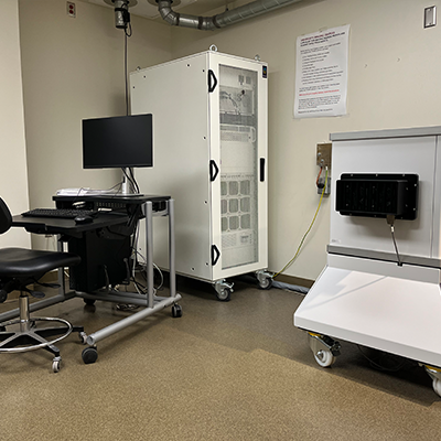
M5 MRI
The M5 compact 1.0 Tesla, high-performance MRI system is designed with a larger bore opening for dedicated imaging of both mice and rats. The system provides non-invasive 3D anatomical, functional, and molecular read-outs in rodents and ex vivo samples.
The M compact, high-performance MRI system enables high-resolution 3D whole-body morphological imaging of mouse models of disease. Biologists and researchers without prior MR or imaging experience can now leverage the gold standard modality – MRI – for morphological imaging. The M5 quickly generates reproducible, quantitative results of phenotypes and therapeutic efficacy. For more information, see the Aspect Imaging M5 website.

Wildtype mouse vertebral column, T2 weighted image obtained with the Aspect Imaging M5 Magnetic Resonance Imager. Yellow arrows indicate clearly defined intervertebral discs (IVDs); magenta arrows indicate fused lumbar IVDs, a normal developmental process. Scale bar = 1 mm. Image courtesy of Dr. Scott Thompson; laboratory of Dr. Laura Stone, Department of Anesthesiology, University of Minnesota.
Applications
- Cancer
- Cardiovascular
- Contrast-based Molecular Imaging
- Diabetes and Obesity
- Embryology/Developmental Biology
- Nephrology, Neurobiology
- Stem Cell/Cell Tracking
Gradients
- Strength– 430 (X,Y,Z) mT/m @ 60 A
- Linearity (for Entire Imaging Volume)– Better than 5%
- Slew Rate (T/m/s)– 1720 T/m/s @ 60 A / 250 us
Imaging Pulse Sequences
| Sequence | Description |
|---|---|
| Spin Echo | 2D DWI – with diffusion gradients IR – with preceding inversion pulse |
| Fast Spin Echo | 2D and 3D DWI – with diffusion gradients IR – with preceding inversion pulse |
| Gradient Echo | 2D and 3D |
| IRSnap | GRE based with multiple inversion time |
| DCE | GRE based Dynamic Contrast Enhancement |
| Cardiac Cine | GRE based ECG triggered Cine |
| DTI | FSE based diffusion tensor imaging |
| Dixon | FSE based fat separation |
| Field of View | 90 x 90 x 60 mm3 spheroid |