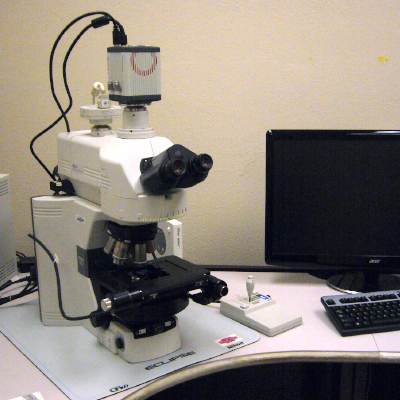
Light Microscopes
Cancer and Cardiovascular Research Building (CCRB)
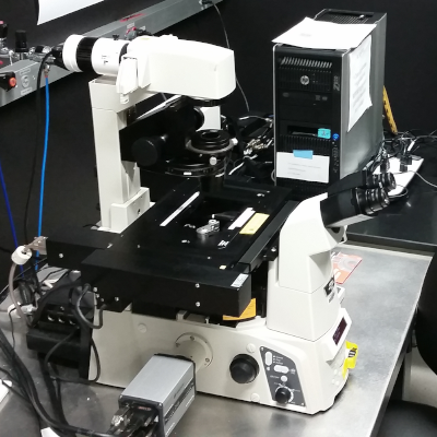
Nikon AX R with NSPARC Super Resolution

Nikon Ti2
Fully motorized (multi-position nosepiece, filters, condenser, z-motor, x/y stage) inverted microscope with 25 mm field of view. This FAST resonant (15 fps full 2K FOV with up to 720 fps at 2048x16 pixels) or galvanomic scanners (up to 8192x8192 pixels for Nyquist resolution at lower magnifications and the larger FOV). This scope has Nikon’s Perfect Focus System.
NSPARC
Next Generation Super-Resolution. The Nikon Spatial Array Confocal (NSPARC) detector utilizes an ultra-low noise photon counting detector array that can collect an image at each scanned point. Final resolutions of < 100 nm are possible using any of the 8 laser lines on the AX R confocal system.
Lasers and detection filters
408, 445, 488, 514, 561, 594, 640 and 730nm excitation lines allowing for multiplexed imaging of many more fluorescent reporters.
Detector
Four channel GaAsP PMT detector modules, including spectral detection along plus Differential Interference Contrast (DIC) transmitted light.
Objectives
4x, 10x, 20x, 20XW, 40x oil, and 60x oil immersion objectives. The system is compatible with many of the UIC’s extensive collection of additional Nikon objectives.
Live cell imaging
Tokai-Hit environmental controls (temp, humidity, CO2).
Software
Nikon Element AI software has built-in multidimensional (multi-XY,Z,T, multichannel) experiment capabilities, including AI analysis and image enhancement tools. The JOBS module allows customization in setting up non-orthogonal experiments with multiple paths and dimensions. Analysis of data can be done even in real-time during the experiment, and the direction of the experiment can even be changed based on the results of the analysis. Fully automated collection and analysis capabilities with JOBS. The UIC staff is excited to help train you from the most basic image acquisition to the full complement of our instruments capabilities.
Nikon C2+ Upright Spectral Confocal and Widefield Imaging System

Nikon Ni
Features include Nikon’s motorized Ni microscope body. The CFI60 objectives for the Ni system are 10x, 20x, 40x oil, and 60x oil, all with DIC capabilities. An LED illumination system for fluorescence, and filters for widefield using DAPI, GFP, and Tex Red filters. There is a Prior motorized x/y stage and color camera for tiling of large areas of interest in brighfield, DIC, fluorescence, and confocal modes.
Laser lines
There are 405 nm, 488 nm, 561 nm, and 640 nm laser lines for excitation
Detectors
Dual GaSP detectors with continuous filters for spectral imaging
Three photo-multiplier tubes (PMTs), plus transmitted light detectors, Nikon DS-U3 color camera
Transmitted light filters
ND2, ND4, ND8, ND16, Green, and Cool Blue
Widefield image capture
Tiling in brightfield, DIC, DAPI, AF488, AF568 and CY5 fluorescence
Includes Elements modules for 2D and 3D Tracking and Measurements, Ratio and FRET imaging
Objectives
|
Magnification |
NA |
Correction |
Optic Properties |
Tube Length/Cover Glass Thickness |
Working Distance (mm) |
Immersion Medium |
|---|---|---|---|---|---|---|
|
4x |
0.13 |
PlanFluor |
PhL DL |
∞/1.2 |
16.5 |
Air |
|
10x |
0.45 |
PlanApo λ |
DIC N1 |
∞/0.17 |
4.0 |
Air |
|
20x |
0.75 |
PlanApo λ |
DIC N2 |
∞/0.17 |
1.0 |
Air |
|
40x |
1.30 |
PlanFluor |
DIC N2 |
∞/0.17 |
0.24 |
Oil |
|
60x |
1.40 |
PlanApo |
DIC N2 |
∞/0.17 |
0.14 |
Oil |
|
100x |
1.40 |
PlanApo VC |
DIC N2 |
∞/0.17 |
0.13 |
Oil |
Nikon Ti-E Inverted Deconvolution Microscope
Nikon Ti-E
The Nikon Ti-E Motorized Microscope with Ibidi live cell environment chamber features include Nikon’s patented PFS Perfect Focus III focus lock technology. 100 Watt Illumination system for transmitted and LED-reflected light applications. Fluorescence accessories for widefield applications for DAPI, GFP, CFP, YFP, RFP and Cy5...and many others.
Light source
SPECTRA III LED light source lines (nm): 390, 440, 475, 510, 555, 594, 639, 748
Software
Nikon Elements including JOBS, deconvolution, time measurements, automated collection of slides, dishes, and plates.
Resources
Live cell imaging
- Live cell imaging reagents
- Ibidi Environmental Stage with CO2 and O2 regulation
- Ibidi dishes and chambered slides
- We also have the Braintree Scientific Automated Syringe pump to perfuse solutions! Holds 6 syringes up to 50cc.
Objectives
New Lambda coat anti-reflective coated objectives at 4x, 10x, 20x, 40x* NAMC Contrast, 60x, and 100x DIC magnifications (*correction collars).
| Magnification | NA | Correction | Optic Properties | Tube Length/Cover Glass Thickness | Working Distance (mm) | Immersion Medium |
|---|---|---|---|---|---|---|
| 4x | 0.13 | PlanFluor | -- | ∞/- | 17.1 | Air |
| 10x | 0.45 | PlanApo | DIC N1 | ∞/0.17 | 4.0 | Air |
| 20x | 0.75 | PlanApo | DIC M/N2 | ∞/0.17 | 1.0 | Air |
| 40x | 0.95 | Plan Apo | DIC MN2, Correction Collar | ∞/0.11-0.23 | 0.14 | Air |
| 60x* | 1.20 | Plan Apo VC with correction collar | ∞/0.15-0.18 | 0.27 | Water | |
| 60x | 1.4 | PlanApo | DIC N2 | ∞/0.17 | 0.13 | Oil |
| 100x | 1.45 | PlanApo | DIC N2 | ∞/0.17 | 0.13 | Oil |
Nikon AZ100M Macro Fluorescence Microscope

Nikon AZ100 Multizoom
Motorized X/Y stage for capturing multi-position or panoramic images, combined with motorized Z-axis for z-stack acquisition, enabling 3D imaging and extended depth-of-field.
Detectors
Photometrics ES2 greyscale 12-bit camera for fluorescence
Nikon DS-Ri1 color 12-bit camera up to 4076x3116 pixels for full-color imaging
Filters
Fluorescence (UV, CFP, GFP, YFP, dsRed, and CY5 filter sets ) and transmitted light with DIC
Software
Nikon Elements AR software for multi-dimensional data collection
Objectives
1X to 8X Optical Zoom Range. Total Magnification: 10x to 400x
| Magnification | NA | Correction | Working Distance (mm) | Immersion Medium |
|---|---|---|---|---|
| 1x | 0.1 | AZ PlanApo | 35 mm | Air |
| 4x | 0.4 | AZ PlanApo | 20 mm | Air |
Filters
| Filter | Excitation Filter (nm) | Dichroic Mirror (nm) | Emission Filter (nm) |
|---|---|---|---|
| DAPI | 360-380 | 400 | 415 (LP) |
| CFP | 426-446 (436 CWL) | 455 LP | 460-500 (480 CWL) |
| YFP | 490-510 (500 CWL) | 515 LP | 520-550 (535 CWL) |
| GFP | 470- | 495 LP | 525- |
| dsRed | 510-555 | 560 LP | 575-595 (580 CWL) |
Leica LMD6500 Laser Dissection Microscope
Leica LMD6500
Laser microdissection uses a microscope to visualize individual cells or cell clusters. Regions of interest are selected by software, excised from the surrounding tissue by a laser, and collected for subsequent molecular analysis.
- Training video using the LMD6500 on JOVE
- LDM works best when using PEN membrane slides. The PEN membrane on glass slides are here
- Videos on the LDM technique can be found here
Transmitted Light Filters
ND2, ND4, ND8, and ND16, Green and Cool Blue
Laser lines
Wavelength: 355 nm, Max. pulse energy: 70 µJ, Pulse frequency: 80 Hz
Detector
LMD CC7000 color camera
Objectives
| Magnification | NA | Correction | Optic Properties | Tube Length/Cover Glass Thickness | Working Distance (mm) | Immersion Medium |
|---|---|---|---|---|---|---|
| 1.25 | 0.04 | HCX FL FLUOTAR | ∞/-/^ | 3.7 | ||
| 5x | 0.12 | UVI | ∞/- | 11.7 | Air | |
| 10x | 0.3 | HCX FL FLUOTAR | Ph2 | ∞/-/D | 11 | Air |
| 20x | 0.4 | HCX FL FLUOTAR | Ph1 | ∞/0-2/C | 6.9 | Air |
| 40x | 0.6 | HCX FL FLUOTAR | Corr XT | ∞/0-2/C | 3.3-1.9 | Air |
| 63x | 0.7 | HCX FL FLUOTAR | Ph2 | ∞/0.1-1.3/C | 2.6 | Air |
Filters
| Filter | Excitation Filter (nm) | Dichoric Mirror (nm) | Emission Filter (nm) | Filter Wheel Position |
|---|---|---|---|---|
| DAPI | 360-370 (365 CWL) | 380 LP | 420 LP | Open |
| U/B/G (triple DAPI/GFP/dsRed) | 385-415 (405 CWL) | 410-470 (440 CWL) | 450-470 (460 CWL) | UV/B/G |
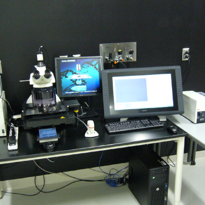
Leica MZ FL III Fluorescence Stereomicroscope
Leica MZ FL III
Sort, manipulate, and image your specimens with the stereo objective in 3D, with large fields of view, large working distances, and an outstanding depth of field. Simply switch to the HR (High Resolution) micro-objective and analyze the quality of the gene expression immediately and without delay via an optical light microscope at a resolution of 0.7 µm (1320 pairs of lines/mm) and an 800x magnification.
Detector
Leica DFC420 C camera. Digital 5 MP color camera with Peltier cooling system to capture high-resolution images for analysis, documentation, and presentation.
Objectives
| Magnification | NA | Correction | Optic Properties | Tube Length/Cover Glass Thickness | Working Distance (mm) |
|---|---|---|---|---|---|
| 0.5x | Plan | 139 | Air | ||
| 1x | 0.035 | PlanApo | -/- | 55 | Air |
| 2x | 0.07 | PlanApo | -/- | 15 | Air |
Filters
| Filter | Wavelengths | |
|---|---|---|
| Excitation Filter (nm) | Emission Filter (nm) | |
| UV | D350/50x | E420 1pv2 |
| GFP1 | ET425/60x | ET505/40m |
| GFP2 | 480/40 | 510 |
| G | 546/10 | 590 |
Jackson Hall
Nikon AX R

Nikon Ti2
Fully motorized (multi-position nosepiece, filters, condenser, z-motor, x/y stage) inverted microscope with 25 mm field of view. This FAST resonant (15 fps full 2K FOV with up to 720 fps at 2048x16 pixels) or galvanomic scanners (up to 8192x8192 pixels for Nyquist resolution at lower magnifications and the larger FOV). This scope has Nikon’s Perfect Focus System.
Live Cell Imaging
Ibidi stage top incubator and environmental controls enabling researchers to perform live cell imaging in plates, slides and dishes
LED light source excitation
390, 440, 475, 510, 555, 575, 637, 748 nm
Widefield camera
Teledyne Photometrics Prime 95B sCMOS
Confocal Imaging lasers and detection filters
408, 488, 561 and 640 nm excitation lines
Widefield Emission Filters
Widefield illumination and filters for a variety of fluorochromes including the Opal dyes UV, CFP, YFP, GFP, dsRed, TRITC, CY5 imaging
| Filter Name | Peak (nm) | Range (nm) |
|---|---|---|
| DAPI | 432 | 36 |
| Opal 520 | 515 | 30 |
| Opal 540 | 544 | 24 |
| Opal 570 | 595 | 31 |
| Opal 620 | 641 | 75 |
| Opal 690 | 680 | 42 |
Objectives
| Magnification | NA | WD (mm) | Immersion Medium |
|---|---|---|---|
| 2x | .10 | 8.5 | Air |
| 4x | .1 | 16.5 | Air |
| 10x | .3 | 15.2 | Air |
| 20x | .7 | 2.3-1.3 | Air |
| 20x LWD* | .95 | 0.23-0.11 | Water |
| 40x | .95 | 0.99-0.9 | Air |
| 40x ELWD* | .6 | 3.6-2.8 | Air |
| 60x | 1.4 | 0.13 | Oil |
| 100x* | 1.49 | 0.13 | Oil |
| 100x* | 1.35 | 0.31-0.28 | Silicone |
| *(available by request) | |||
Nikon A1Rsi HD Confocal with SIM Super Resolution

Nikon Ti-E
Nikon Ti-E Motorized Microscope featuring PFS Perfect Focus III focus lock technology. LED Illumination systems for both transmitted and fluorescence light applications. Fluorescence accessories for widefield applications for DAPI, GFP, RFP, and Cy5.
SIM Structured Illumination System-Using high-frequency Structured Illumination, the Nikon N-SIM can achieve an image resolution of 120 nm. With a temporal resolution of up to 0.6 sec/frame, N-SIM enables super-resolution time-lapse imaging capture of dynamic molecular interactions in living cells.
Laser lines
(A1R): 405 nm, 488 nm, 561 nm, 640 nm
Lasers (N-SIM): 405 nm, 488, nm, 561 nm, 640 nm
Detectors
The A1R has four PMTs for confocal detection, two of them GaAsPs, with double the sensitivity of traditional PMTs. There is also a transmitted light detector and a 32-channel spectral detector.
N-SIM: Hamamatsu FLASH 4
Software
The Nikon Elements software has the JOBS package that can automate image collection for high-content screening of plates.
Objectives
|
Magnification |
NA |
Correction |
Optic Properties |
Tube Length/Cover Glass Thickness |
Working Distance (mm) |
Immersion |
|---|---|---|---|---|---|---|
|
2x |
0.1 |
PlanApo |
8.5 mm |
Dry |
||
|
10x |
0.45 |
PlanApo |
DIC N1 |
∞/0.17 |
4 |
Dry |
|
20x |
0.75 |
PlanApo VC |
DIC N2 |
∞/0.17 |
1 |
Dry |
|
40x |
0.6 |
S PlanFluor |
DIC N1, Correction Collar |
∞/0-2 |
3.6 |
Dry |
|
40x |
0.95 |
PlanApo |
DIC MN 2, Correction collar |
∞/0.11-0.23 |
0.14 |
Dry |
|
60x |
1.27 |
PlanApo IR with correction collar |
DIC N2 |
∞/0.15-0.19 |
0.18-0.16 |
Water |
|
60x |
1.4 |
Plan Apo Lambda |
DIC N2 |
∞/0.17 |
0.13 |
Oil |
|
100x |
1.49 |
SR ApoTIRF with correction collar |
DIC N2 |
∞/0.13 |
0.12 |
Oil |
Filters - Confocal
| Filter cube | Emission band | Common dyes |
|---|---|---|
| 99020 | 450/40 | DAPI |
| 99022 | 525/50 | GFP, AF488 |
| 99023 | 600/50 | mCherry, AF568 |
| 99023 | 660LP | AF647, Cy5 |
Filters - Camera
| Filter cube | Availability | Excitation band | Dichroic mirror | Emission band | Common dyes |
|---|---|---|---|---|---|
| Chroma 49028 | Installed | 395/25 | T425lprx | 460/50 | DAPI |
| Chroma 49002 | Installed | 470/40 | T495lprx | 525/50 | GFP, AF488 |
| Chroma 49005 | Installed | 545/30 | T570lp | 620/60 | dsRed, AF568 |
| Chroma 49006 | Installed | 620/60 | T660lprx | 700/75 | AF647, Cy5 |
| Chroma 49303 | Available on request | 495/25 | T515lprx | 537/29 | Opal 520 |
| Chroma 49304 | Available on request | 546/10 | T556lprx | 572/23 | Opal 570 |
| Chroma 49306 | Available on request | 580/25 | T600lprx | 615/30 | Opal 620 |
| Opal 620 | Available on request | 590/20 | 635/75 | Opal 620 | |
| Chroma 49022 | Available on request | 650/45 | T685lprx | 720/60 | Opal 690, Cy5.5 |
Filters - SIM
| Filter cube | Availability | Excitation band | Dichroic mirror | Emission band | Effective emission window | Common dyes |
|---|---|---|---|---|---|---|
| SIM405 | Installed | 395/20 | 440LP | 460/50 | 440-485 | DAPI |
| SIM488 | Installed | 480/20 | 515LP | 523/45 | 515-545 | GFP, AF488 |
| SIM561 | Installed | 561/10 | 590LP | 605/70 | 590-640 | mCherry, AF568 |
| SIM640 | Installed | 620/60 | 665LP | 700/75 | 665-738 | AF647, Cy5 |
Nikon A1R HD Multiphoton Microscope

Nikon FN1
The Nikon A1RMP confocal is connected to an upright Nikon FN1 microscope featuring a Plan Apo LWD 25x water immersion, NA 1.1 objective most suitable for imaging deep (it has a 2 mm working distance) into a fixed or living sample. In addition to laser scanning confocal imaging, conventional, and resonant visible laser confocal imaging, a Spectra-Physics 15W Mai Tai eHP tunable IR laser and six, three GaAsPs (epi direction) and three conventional PMTs (trans direction) non-descanned GaAsP detectors allow multiphoton visualization of multiple fluorophores located deep within the specimen. The system has both galvonomic (4K) and resonant (1k, 8Khz, up to 720fps) scanners that can be used in concert simultaneously for photoactivation and MP imaging. A encoded Prior motorized stage and 200 µm Piezo objective motor can be used for tiling or multiple-position imaging.
The system is also equipped with Second Harmonic Generation (SHG) detection. SHG doesn't involve the excitation of molecules like other techniques such as fluorescence microscopy.
This instrument was funded in 2012 by the Minnesota Partnership in conjunction with the Mayo Clinic Foundation, Rochester, MN.
Laser lines
405, 457, 488, 514, 561 and 637 nm
Objectives
|
Magnification |
NA |
Correction |
Optic Properties |
Tube Length/Cover Glass Thickness |
Working Distance (mm) |
Immersion |
|---|---|---|---|---|---|---|
|
10x |
0.3 |
PlanFluor |
DIC L |
∞/0.17 |
16 |
Dry |
|
10x |
0.3 |
Fluor |
DIC L |
∞/0 |
2 |
Water |
|
10x |
0.5 |
Plan Apo |
Correction Collar |
∞/- |
5.5 |
Glycerol |
|
20x |
0.45 |
PlanFluor |
DIC L |
∞/0-2 |
7.4 |
Dry |
|
20x |
0.75 |
PlanApo lambda |
DIC N2 |
∞/0.17 |
1 |
Dry |
|
25x |
1.1 |
ApoLWD |
DIC 2, Correction Collar |
∞/0-0.17 |
2 |
Water |
|
40x |
0.8 |
Fluor |
DIC M |
∞/0 |
2 |
Water |
|
100x |
1.3 |
PlanFluor |
|
∞/0.17 |
0.2 |
Oil |
Filters - Multiphoton
| Filter cube | Emission band | Common dyes |
|---|---|---|
| 99020 | 450/40 | DAPI |
| 9920-IR | 460/50 | DAPI |
| 99022 | 525/50 | GFP, AF488 |
| 99021 | 485/30 | CFP |
| 99023 | 600/50 | mCherry, AF568 |
| 99023 | 660LP | AF647, Cy5 |
| 99024 | 545/40 | YFP |
Filters - MP Non-Descanned Detector (NDD)
| Filter cube | Availability | NDD Type | Emission band | Common dyes |
|---|---|---|---|---|
| 99124 | Installed | Epi | 425/50 |
SHG collagen, DAPI TPE |
| 99121 | Installed | Epi | 510/80 | GFP |
| 99121 | Installed | Epi NDD | 605/70 | mCherry |
| 99020 | Installed | Trans | 450/40 |
SHG collagen, DAPI TPE |
| 99121 | Installed | Trans | 515/70 | GFP |
| 99121 | Installed | Trans | 605/70 | mCherry |
| 99321 | Available on request | 440/80 |
SHG collagen, DAPI TPE |
|
| 99324 | Available on request | 540/80 | YFP | |
| 99324 | Available on request | 620/52 | mCherry |
3i AxL Cleared Tissue LightSheet

Cleared Tissue LightSheet
The AxL Cleared Tissue LightSheet (AxL CTLS) is an advanced, fully automated macro zoom imaging system designed for large-scale, high-resolution visualization of whole organs and small animals. It features high-NA apochromatic objectives, dual-sided light sheet illumination, and custom excitation optics. Leveraging patented axially swept light sheet microscopy (ASLM), it delivers an exceptionally thin, extended, and uniform light sheet for large-scale high-resolution imaging.
Lasers
488 nm, 514nm, 561 nm, 638 nm
Detector
Hamamatsu Orca Fusion BT
Objectives
|
Magnification |
NA |
Correction |
Working Distance (mm) |
|---|---|---|---|
| 1.0 | 0.25 | Plan Neofluar | 56 |
|
1.5 |
0.37 |
Apo Z |
30 |
Sartorius Incucyte SX5
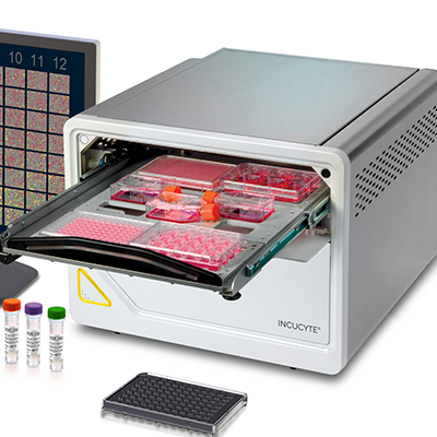
Incucyte SX5
The Incucyte SX5 Live-Cell Analysis System is a real-time cell monitoring and surveillance system that automatically captures and analyzes images of living cells around-the-clock for days, weeks, or months, while cells remain undisturbed inside a standard tissue culture incubator.
Vessel Compatibility
Your cells must be in a compatible vessel to utilize the Incucyte SX5. Please review the list below to see if your vessel is compatible.
UIC Incucyte SX5 compatible vessels
System Features
Available Modules
- Image Lock - Enables precise, repeat imaging of the same field of view, allowing for cell motility studies as well as time-lapse image registration required for precise movie generation.
- Scratch Wound - Automated detection and quantification of wounds from each well of a 96-well plate in order to analyze cell migration and invasion with or without labels. The integrated analysis algorithm automatically masks each image to identify the position of the wounded (cell-free) and unwounded (cell-occupied) zones, to deliver robust measurements of wound width, wound confluence and relative wound density (RWD) for the entire time-course of the experiment.
- Whole Well - Acquire whole well images of select multi-well and 35mm dishes to enable monitoring and analysis of rare events within a cell population.
- Dilution Cloning - Automatically identify wells containing clones and track colony formation over time. Enable whole well images in 96- ad 384-well microplates.
- Spheroid - Analyze growth, viability, and invasion of single spheroids in round-bottom, multi-well formats or monitor multiple spheroids in flat-bottom plates. Enables kinetic, label-free or fluorescence-based analysis in physiologically relevant conditions with minimal disruption. Automatically quantifies spheroid size, shape, count, and invasive spread over time.
LUMICKS C-Trap Optical Tweezers

The University Imaging Centers (UIC) is excited to announce the installation of the Lumicks C‑Trap Optical Tweezers at UIC Jackson Hall, made possible through an awarded NIH S10 collaboration with Dr. Wendy Gordon from the Medical School’s Department of Biochemistry, Molecular Biology, and Biophysics.
The C-Trap is a dynamic single-molecule imaging system equipped with 488, 561, and 638 nm lasers that integrates optical tweezers with multicolor fluorescence microscopy and microfluidics. It is designed to simultaneously manipulate biomolecules and visualize their interactions in real-time with sub-piconewton force sensitivity and nanometer spatial precision.
The C-Trap is used across diverse biophysical and biomedical research fields to study:
- Molecular Motors: Tracking the stepping mechanics, velocity, and stall forces of motors such as kinesin and myosin along cytoskeletal filaments.
- Protein Folding and Mechanics: Measuring the forces required for protein unfolding/folding and observing conformational changes using techniques like FRET.
- Cell Mechanics and Virology: Measuring cell membrane stiffness, mechanosensing, and the mechanisms of viral assembly or entry into host cells.
- DNA and RNA Interactions: Visualizing DNA-binding proteins (e.g., Cas9, polymerases) as they move, bind, or repair DNA under varying mechanical tension.
Chromatin Organization: Following the dynamic movement of chromatin remodelers, nucleosome sliding, and histone eviction.
Snyder Hall
Nikon AX R

Nikon NiE
Nikon NiE fully motorized (multi-position nosepiece, filters, condenser, z-motor, x/y stage) upright microscope with 25 mm field of view. This resonant (15 fps full FOV with up to 720 fps at 2048x16 pixels) and galvanomic scanners (up to 8192x8192 pixels for Nyquist resolution at lower magnifications or larger FOVs).
Lasers and detection filters
408, 445, 488, 514, 561, and 640 nm excitation lines allow for multiplexed imaging of DAPI, CFP, YFP, GFP, FITC, dsRed, TRITC, CY5, and many more fluorescent reporters.
Detector
Four-channel GaAsP PMT detector modules, including spectral detection along with transmitted light.
Objectives
2x, 4x, 10x, 20x, 40x, 60x, 100x including water, silicon and oil immersion options.
Equipped for Differential Interference Contrast (DIC)
Software
Nikon Element ER software has built-in multidimensional (multi-XY, Z, T, multichannel) experiment capabilities. The JOBS module allows customization in setting up non-orthogonal experiments with multiple paths and dimensions. Analysis of data can be done in real-time during the experiment, and the direction of the experiment can even be changed based on the results of the analysis. Fully automated collection and analysis capabilities with JOBS.
Nikon A1si Confocal and Widefield Microscope

Nikon Eclipse Ti2
The A1si system is mounted on a Nikon Eclipse Ti2 inverted scope equipped with Perfect Focus System, fluorescence filters with DIC optics and a widefield Hamamatsu Flash 4 camera. The A1si's highest pixel resolution mode is up to 4096 x 4096 pixels.
Laser lines
405 nm diode at 38mW, Argon multiline laser 457/477/488/514nm at 40mW, 561 nm diode at 40mW, 638 nm diode at 10mW
Detectors
4 PMTs, a Transmitted light detector, and a 32-channel spectral detector
Live cell imaging
There is also an IBIDI environmental stage top incubator system for long-term live cell imaging.
Software
Nikon NIS Elements imaging software is used to control acquisition and analysis. Nikon JOBS will permit investigators to create complex automated image acquisitions with intelligent analysis.
Objectives
|
Magnification |
NA |
Correction |
Optic Properties |
Tube Length/Cover Glass Thickness |
Working Distance (mm) |
Immersion Medium |
|---|---|---|---|---|---|---|
|
4x |
0.2 |
Plan Apo |
-- |
∞/-- |
20 |
Air |
|
10x |
0.3 |
Plan Apo |
-- |
∞/0.17 |
4 |
Air |
|
20x |
0.75 |
Plan Apo |
DIC N2 |
∞/0.17 |
1 |
Air |
|
60x |
1.2 |
Plan Apo |
DIC N2, Correction collar |
∞/0.15-0.18 |
0.27 |
Water |
|
60x |
1.4 |
Plan Apo |
DIC H |
∞/0.17 |
0.21 |
Oil |
Widefield Filters
|
Common Fluorophore |
Excitation Filter (nm) | Dichroic Mirror (nm) | Emission Filter (nm) |
|---|---|---|---|
|
DAPI |
340-380 | 400LP | 435-485 |
|
CFP |
426-446 | 455LP | 460-500 |
|
GFP |
465-495 | 505LP | 515-555 |
|
YFP |
490-515 | 520LP | 520-560 |
| Texas Red | 532-587 | 595LP | 608-683 |
Confocal Filters
|
Common Fluorophore |
Excitation Filter (nm) | Dichroic Mirror (nm) | Emission Filter (nm) |
|---|---|---|---|
|
DAPI |
DS350/50 | BS20/80 | 460/50 |
|
CFP |
DS440/40 | 400-457/514 | 470/40 |
|
GFP |
D480/30 | DM405/488 | D535/40 |
|
YFP |
DS 510/50 | 400-457/514 | D 555/30 |
| dsRed | DS540/25 | 408/488/543 | D600/50 |
| Cy5 | DS640/20 | DM458/515 | D685/70 |
Nikon 90i
Nikon Eclipse 90i
The Nikon Eclipse 90i is a fully motorized upright microscope configured to image in transmitted light, DIC, and epi-fluorescence modes. Fluorescence cubes are available for DAPI, GFP, RFP, and HYQ Cy5. Fluorescence illumination is provided by a Lumencor Sola Light Engine. You can do timelapse, XYZ position, Z stacks, multiple excitations or emission wavelengths, and tiling experiments. Equipped with a Hamamatsu Orca Flash 4.0 v2 CMOS monochrome camera and a Nikon D2-Fi2 color camera running on Nikon Elements software.
Transmitted Light Filters
ND8, ND32, and NCB11
Light engine
Lumencor Sola Light Engine
Detectors
This instrument has two cameras:
- Hamamatsu Orca Flash 4.0 v2 (CMOS) monochromatic Camera.
- Nikon Color DS-Fi2 CCD Camera.
Objectives
| Magnification | NA | Correction | Optic Properties | Tube Length/Cover Glass Thickness | Working Distance (mm) | Immersion Medium |
|---|---|---|---|---|---|---|
| 4x | 0.13 | Plan Fluor | -- | ∞/- | 17.1 | Air |
| 10x | 0.45 | Plan Apo | ∞/0.17 | 4 | Air | |
| 20x | 0.75 | Plan Apo | DIC N2 | ∞/0.17 | 1 | Air |
| 40x | 0.75 | Plan Fluor | DIC M | ∞/0.17 | 0.72 | Air |
| 100x | 1.4 | Plan Apo VC | DIC N2 | ∞/0.17 | 0.21 | Oil |
| 100x | 1.3 | Plan Fluor | Ph3 DLL | ∞/0.17 | 0.2 | Oil |
Filters
| Filter | Excitation Filter (nm) | Dichroic Mirror (nm) | Emission Filter (nm) |
|---|---|---|---|
| DAPI | 325-375 (350 CWL) | 400 LP | 435-485 (460 CWL) |
| DAPI/FITC/TRITC/Cy5 (Quad) | 380-410 (395 CWL) | 410-475 (443 CWL) | 425-465 (445 CWL) |
| DAPI/FITC/TRITC/Cy5 (Quad) | 475-500 (488 CWL) | 505-545 (525 CWL) | 505-535 (520 CWL) |
| DAPI/FITC/TRITC/Cy5 (Quad) | 540-565 (553 CWL) | 575-625 (690 CWL) | 575-620 (598 CWL) |
| DAPI/FITC/TRITC/Cy5 (Quad) | 625-650 (638 CWL) | 655-725 (690 CWL) | 670-740 (705 CWL) |
| GFP | 450-490 (470 CWL) | 495 LP | 500-550 (525 CWL) |
| DS RED | 530-560 (545 CWL) | 570 LP | 590-650 (620 CWL) |
| Cy5 | 590-650 (620 CWL) | 660 LP | 663-738 (700 CWL) |
Nikon AZ100 C1si Spectral Confocal Microscope

Nikon AZ100 Multizoom
The AZ100 Multizoom Macroscope with the C1si Spectral Confocal attachment makes the ultimate platform for in vivo imaging. Ideal for plant biology, developmental biology, cell biology, stem cell, and tissue research, the AZ-C1si allows the researcher to view large specimens in confocal mode. The AZ-C1si can capture fields of view up to 1cm and permits deeper confocal imaging than conventional microscopes thanks to its large working distance objectives. Whole organisms can be monitored and documented over time (for example, embryos) offering a wealth of continuous information on development or the organism’s response to experimental variables. Both conventional PMTs and Spectral detectors are installed on this instrument.
Laser lines
405 nm, 457 nm, 488 nm, 514 nm, 567 nm, 637 nm for image, co-localization, FRAP, FRET, and spectral studies.
Objectives
The system is equipped with 1x, 4x, and 5x objectives with variable optical zoom.
|
Magnification |
NA |
Correction |
Working Distance (mm) |
Immersion Medium |
|---|---|---|---|---|
|
1x |
0.1 |
AZ Plan Apo |
35 mm |
Air |
|
4x |
0.4 |
AZ Plan Apo |
20 mm |
Air |
|
5x |
0.5 |
AZ Plan Fluor |
15 mm |
Air |
Filters
|
Filter |
Excitation Filter (nm) |
Dichroic Mirror (nm) |
Emission Filter (nm) |
|---|---|---|---|
|
DAPI |
D350/50 |
BS20/80 |
460/50 |
|
GFP |
D480/30 |
DM405/488 |
D535/40 |
|
TRITC |
D540/25 |
405/488/543 |
D605/55 |
|
Cy5 |
D640/20 |
DM458/515 |
D680/30 |
Channels
|
Channels |
Dichroic Mirror (nm) |
Barrier Filters (nm) |
|---|---|---|
|
Channel 1 |
SDM560, SDM510, SDM490 |
2nm Spectral Monochromator Grating |
|
Channel 2 |
SDM640, SDM560 |
2nm Spectral Monochromator Grating |
|
Channel 3 |
SDM640 |
BA560-620, BA560-660, BA655-755 |
Molecular and Cellular Biology (MCB)
Leica Stellaris 8

Leica DMi8
Inverted laser scanning confocal microscope with both galvonomic and resonance scanners and 5 spectral internal HyD-S detectors.
Laser lines
A fixed 405nm violet laser and pulsed white-light laser (WLL) that is tunable from 440-790nm allows for precise excitation control and multi-spectral imaging.
Detector
The WLL and HyD-S detectors allow for Leica's patented Tau gating and separation modalities using fluorescence lifetime measurements.
Live cell imaging
Environmental control enables long-term live cell imaging and the Leica Navigator interface streamlines stage control and multi-position high-speed tiling.
Objectives
20x/0.7NA multi-immersion, 40x 1.25NA glycerol motCORR, 63x 1.4NA oil, and 63x 1.2NA water.