Molecular Biology Imagers
Jackson Hall
Typhoon FLA 9500
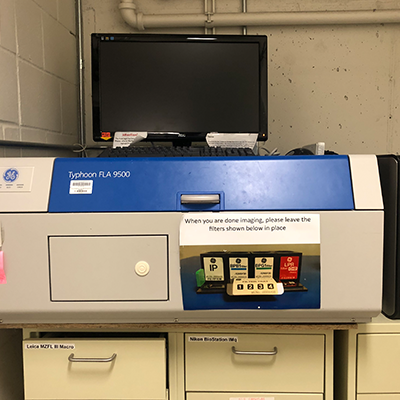
Typhoon FLA 9500 is a variable mode laser fluorescent and phosphor scanner with modular access to the optical components, providing both versatile and flexible imaging for precise quantitation of proteins, nucleic acids, and other biomolecules.
- Versatility: imaging of multifluorescent-, chemifluorescent-, radioisotope-labeled, and colorimetric samples
- High resolution and quantitation: a pixel resolution of up to 10 µm and a linear signal response over five orders of magnitude provides precise quantitation in gels, blots, tissue sections, and arrays
- High sample throughput: scanning area of 40 × 46 cm enables simultaneous imaging of up to 20 gels or blots, measuring 10 × 8 cm in size. This facilitates comparisons among blots and reduces workload and waiting time
- Flexibility: optimized performance for new applications by adapting the system with stages, detectors, filters, and lasers
- 2-D DIGE imaging: simultaneously image two 2-D DIGE gels for differential expression studies
- Visible and infrared fluorescence imaging: optional near-infrared excitation for imaging IRDye™ and other infrared dyes
The system provides several imaging modes at any time, including fluorescence and chemifluorescence, filmless autoradiography, and digitization of colorimetrically-stained samples (e.g., Coomassie™ blue and silver stain). It is also adequate for quantitative chemiluminescent imaging that does not require maximum sensitivity.
The system is optimized for 2-D DIGE imaging for differential protein expression studies, ECL Plus chemifluorescent imaging for quantitative protein detection by Western blotting, and multifluorescent ECL Plex imaging for precise quantitation of two or more proteins in the same blot.
Typhoon FLA 9500 comes equipped with both bi-alkali and multialkali photomultiplier tubes (PMT). This combination provides excellent detection over a very broad spectrum. Each PMT is selected for optimal response to the detected emission wavelength.
The system accommodates four automated filters. Filters are easily accessed and exchanged without tools for attaining optimal imaging conditions.
Stages give the optimal imaging conditions for a range of samples. Samples include agarose and polyacrylamide gels, membranes, DIGE gels, radioisotope-labeled samples, as well as microplates and glass slides with the TP plug-in. The system can simultaneously scan two DIGE gels, each measuring up to 20 x 25 cm. The stages are easily removed from the system for cleaning.
A fast 1000 μm prescan function gives a rapid overview of the sample. The system provides a linear signal response over five orders of magnitude, providing a suitable basis for precise quantitation.
Lasers can be exchanged in the field to accommodate new applications and fluorophores.
Amersham Imager 600UV
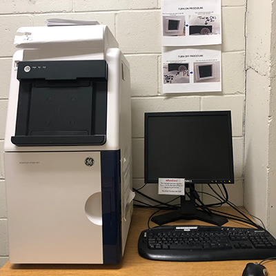
Amersham Imager 600 series is a new range of sensitive and robust imagers for the capture and analysis of high-resolution digital images of protein and DNA samples in gels and membranes. These multipurpose imagers bring high-performance imaging to chemiluminescence, fluorescence, and colorimetric applications. The design of the Amersham Imager 600 combines Western Blotting application expertise with optimized CCD technology and exceptional optics.
The system has integrated analysis software and an intuitive workflow to generate and analyze data quickly and easily.
Snyder Hall
Maestro In-vivo Imaging System
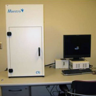
Maestro provides users with a patented method of removing autofluorescence emitted from images of plant and animal tissues to reveal otherwise hard-to-detect labeled targets of interest. The dramatic improvement in signal-to-noise can increase sensitivity up to several hundred-fold, enabling much smaller or fainter signals from biological targets to be detected earlier and accurately measured. The increased sensitivity over standard imaging techniques provided by Maestro’s high-quality spectral imaging technology enables experiments and models which simply cannot be performed on any other system.
The Maestro In vivo imaging system can capture both fluorescent and bioluminescent signals.
Filters: 420 to 720 nm