Electron Microscopes
Sample Preparation Instruments
In addition to electron microscopes, the UIC also provides essential EM sample preparation tools, including a critical point dryer, sputter coater, and ultramicrotome instruments. For more information visit our sample preparation and ancillary equipment page.
Snyder Hall
JEOL JEM-1400Plus Transmission Electron Microscope (TEM)
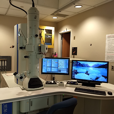
The JEM-1400Plus features high resolution/high contrast imaging, outstanding S/TEM analytical performance, 3D tomography, and montaging. This compact, easy-to-use TEM is suitable for biological, polymer, and materials science applications.
The JEM-1400Plus is equipped as follows:
• 60-120 kV tungsten filament (supports LaB6 filaments)
• Windows-based graphic user interface
• Motorized stage for sample positioning, including image tiling
• Automated tomography tilt-series acquisition
• Bottom-mounted Advanced Microscopy Techniques XR16 camera.
Hitachi S3500N Variable Pressure Scanning Electron Microscope (SEM)
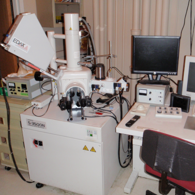
Hitachi S3500N Variable Pressure Scanning Electron Microscope (SEM)
Features include:
- Quartz PCI digital imaging
- Motor-driven X & Y stage
- Environmental Secondary Electron Detector (ESED)
- Absorbed Electron EBIC detector
- Robinson Backscattered Electron Detector (BSE)
- Standard Secondary Electron Detector (SED)
- Infrared ChamberScope
- Windows NT operating system
- Emitech K-1150 Cryogenic System with cryo-prep unit
- Airlock interface to SEM
Serial Block-Face Scanning Electron Microscopy (SBF-SEM)
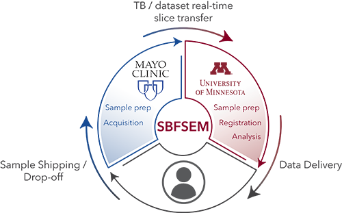
The University Imaging Centers in collaboration with the Mayo Clinic Microscopy and Cell Analysis Core, and the Minnesota Supercomputing Institute provide Minnesota researchers access to serial block face electron microscopy and data sharing, thanks to funding from the Minnesota Partnership for Biotechnology and Medical Genomics.
The award provides electron microscopy instrumentation, cyberinfrastructure and expertise to researchers at Mayo Clinic, the University of Minnesota and the Hormel Institute. This unique microscopy allows investigators to visualize biological structures at high resolution in 3-D space, bridging the gap between super-resolution and multiphoton optical microscopy and standard transmission electron microscopy.
The UIC can prepare your samples and work with you and the staff at the Mayo Clinic Microscopy and Cell Analysis Core to get them in the imaging queue. While we provide consultation for each project, the standard workflow and protocols are available in the PDF documents below.