Sample Preparation & Ancillary Equipment
Snyder Hall
Cressington 108 Auto Sputter Coater
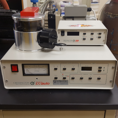
The Cressington 108 Auto Sputter Coater deposits a thin layer of metal (60% gold / 40% palladium) on mounted samples for improved signal in the SEM. The coater is equipped with a rotating-tilting stage for better sample coverage, and a thickness detector for controlled metal deposition.
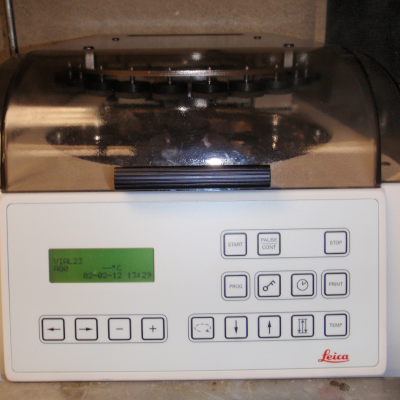
Leica CM1800 Cryostat

Leica CM1800 Cryostat utilizes up-to-date microprocessor technology ensuring easy operation. All functions are protected against dirt and humidity by a plastic foil. It features an easily readable display including an automatic display of errors and a motorized coarse feed which enables rapid trimming and sectioning.
Leica Tissue Processor
Tissues can be processed into a variety of resins for subsequent electron and light microscopy analysis. The pre-heat system allows the correct reagent temperature to be maintained throughout processing. Small vials are available for EM processing and larger for LM which can also be used for high throughput sample processing runs where over 150 EM samples can be processed in one run.
Leica Ultracut UCT Ultramicrotome
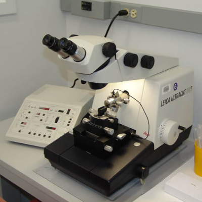
The ultramicrotome is an instrument commonly used to prepare TEM samples. It uses either a glass or diamond knife to cut ultra thin sections of specimens embedded in epoxy. The sections are approximately 60 to 100 nanometers in thickness and 0.5 to 3 mm in width and can be stained and mounted onto TEM grids for viewing. The ultramicrotome is also used to create mirror-smooth surfaces on samples mounted in epoxy, the applications for which include secondary imaging on the SEM and nanoindentation. Preparation on smooth samples for surface analysis need not always be of samples that are embedded, as sometimes bulk will work.
Tousimis Autosamdri-931 Critical Point Dryer

The Tousimis AUTOSAMDRI-931 critical point dryer dehydrates samples prior to examination under the SEM. It uses liquid carbon dioxide at a specific temperature and pressure to significantly reduce drying artifacts due to liquid surface tension. The 931 has a 1.25”-diam, 1.25”-depth chamber with an optional chamber extender. Its default "Automode" setting is suitable for the majority of sample types, and allows you to step away from the dryer. Users can also create and save custom recipes for specific sample needs. Multiple specimen holder sizes are available.
Cancer & Cardiovascular Research Building (CCRB)
PELCO easiSlicer Vibratome

The PELCO vibratome provides exceptional stability and perfect control of section thickness down to 40 microns, with minimal z-axis vibration.
- Precision design for accurate, high quality sections with minimal z-axis vibration
- Novel bath design utilizes unique easy-locking base making it simple to clean
- Anodized specimen tray magnetically locks onto bath
- Optional magnetic base for tissue mounts available
- Cuts sections to 40 microns
- LED lights indicate active vibratory status and cutting window boundaries
- Standard removable blade holder securely holds Feather® blades at a 15º
preset angle; other optional angles available
| Description | Spec |
|---|---|
| Maximum Specimen Size | 40 x 40mm |
| Maximum Specimen Height | 15mm |
| Specimen Bath | Spring-loaded; polypropylene |
| Specimen Tray | Rectangular with magnet release. |
| Blade Type | Feather blades recommended |
| Blade Angle | 15° standard; 18°, 21° available |
| Sectioning Window | 10 - 40mm |
| Specimen Height Adjustment (Z-Axis) | 15mm total travel in 1μm increments; tactile click stops at 5μm |
| Cutting Arm Speed (Y-Axis) | Forward: 0 - 2.0mm/sec Reverse: 5mm/sec Travel: 40mm total |
| Vibratory Amplitude (X-Axis) | 0 - 1.00mm total; continuously adjustable |
| Cutting Arm Frequency | 50Hz ±5 factory set |
| Magnification | 2X magnifier |
| Illumination | LED illumination; LOW/OFF/HIGH |
PELCO BioWave Pro Microwave Processor
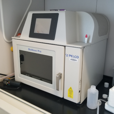
Sample preparation methodology for light and electron microscopy for live and fixed cell and tissue imaging.
- Variable power
- ColdSpot with temperature controlled chiller
- Vacuum chamber
ONIX Microfluidic Perfusion Platform
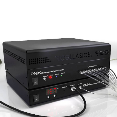
The ONIX Microfluidic Perfusion Platform delivers unprecedented control for live cell imaging experiments and enables dynamic time-lapse experiments.
The UIC stocks a moderate supply of MS04S (mammalian cells), Y04C (yeast haploid cells), B04A (bacterial cells), and C04A (Chlamydomonas cells) plates. Users may purchase their own plates if other designs are desired (gradient, yeast diploid, open top,...).
Countess Automated Cell Counter
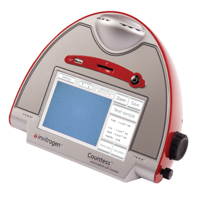
The Countess Automated Cell Counter offers easy and accurate cell counting and viability counts. You can get all the data you need about your cell cultures in just 30 seconds without using a hemocytometer.
The Countess automated cell counter is designed to read samples from 1 × 10⁴ cells/mL to 1 × 10⁷cells/mL, with the highest accuracy between 1 × 10⁵ cells/mL and 4 × 10⁶ cells/mL
- What size cells does it count? - 5 um cells to 60 um cells
- What does it use to distinguish live and dead cells? - Dye exclusion using 0.2% Trypan Blue dye. Live cells exclude the dye. Dead cells take it up into their cytoplasm.
- How does it count the cells? - A sophisticated image analysis algorithm identifies objects, classifies cells by roundness and size and then distinguishes live cells from dead cells by their staining pattern.
- Does it measure cell size? - Yes. It measures the size of both the live cells and the dead cells and reports the average size of each cell population.