Confocal Microscopes
Requesting Training and Services
If you are interested in gaining access to UIC resources or requesting services please follow the steps on our training and service request page.
Cancer and Cardiovascular Research Building (CCRB)
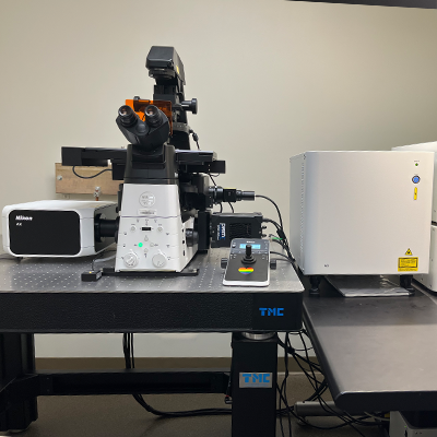
Nikon AX R with NSPARC Super Resolution
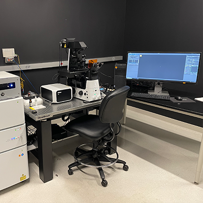
Highlights of the new Nikon AX R / Ti2 with NSPARC include:
NSPARC: Next Generation Super-Resolution. The Nikon Spatial Array Confocal (NSPARC) detector utilizes an ultra-low noise photon counting detector array that can collect an image at each scanned point. Final resolutions of < 100 nm are possible using any of the 8 laser lines on the AX R confocal system.
Nikon Ti2: Fully motorized (multi-position nosepiece, filters, condenser, z-motor, x/y stage) inverted microscope with 25 mm field of view. This FAST resonant (15 fps full 2K FOV with up to 720 fps at 2048x16 pixels) or galvanomic scanners (up to 8192x8192 pixels for Nyquist resolution at lower magnifications and the larger FOV). This scope has Nikon’s Perfect Focus System.
Lasers and detection filters: 408, 445, 488, 514, 561, 594, 640 and 730nm excitation lines allowing for multiplexed imaging of many more fluorescent reporters.
Detector: Four channel GaAsP PMT detector modules, including spectral detection along plus Differential Interference Contrast (DIC) transmitted light.
Objectives: 4x, 10x, 20x, 20XW, 40x oil, and 60x oil immersion objectives. The system is compatible with many of the UIC’s extensive collection of additional Nikon objectives.
Tokai-Hit environmental controls (temp, humidity, CO2) for live cell imaging.
Nikon Element AI software has built-in multidimensional (multi-XY,Z,T, multichannel) experiment capabilities, including AI analysis and image enhancement tools. The JOBS module allows customization in setting up non-orthogonal experiments with multiple paths and dimensions. Analysis of data can be done even in real-time during the experiment, and the direction of the experiment can even be changed based on the results of the analysis. Fully automated collection and analysis capabilities with JOBS. The UIC staff is excited to help train you from the most basic image acquisition to the full complement of our instruments capabilities.
Nikon C2+ Upright Spectral Confocal and Widefield Imaging System
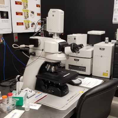
Features include Nikon’s motorized Ni microscope body. The CFI60 objectives for the Ni system are 10x, 20x, 40x oil, and 60x oil, all with DIC capabilities. An LED illumination system for fluorescence, and filters for widefield using DAPI, GFP, and Tex Red filters. There is a Prior motorized x/y stage and color camera for tiling of large areas of interest in brighfield, DIC, fluorescence, and confocal modes.
- There are 405 nm, 488 nm, 561 nm, and 640 nm laser lines for excitation
- Dual GaSP detectors with continuous filters for spectral imaging
- Three photo-multiplier tubes (PMTs), plus transmitted light detectors, Nikon DS-U3 color camera
- Transmitted Light Filters: ND2, ND4, ND8, ND16, Green, and Cool Blue
- Widefield image capture, including tiling in brightfield, DIC, DAPI, AF488, AF568 and CY5 fluorescence
- Includes Elements modules for 2D and 3D Tracking and Measurements, Ratio and FRET imaging
Objectives
|
Magnification |
NA |
Correction |
Optic Properties |
Tube Length/Cover Glass Thickness |
Working Distance (mm) |
Immersion Medium |
|---|---|---|---|---|---|---|
|
4x |
0.13 |
PlanFluor |
PhL DL |
∞/1.2 |
16.5 |
Air |
|
10x |
0.45 |
PlanApo λ |
DIC N1 |
∞/0.17 |
4.0 |
Air |
|
20x |
0.75 |
PlanApo λ |
DIC N2 |
∞/0.17 |
1.0 |
Air |
|
40x |
1.30 |
PlanFluor |
DIC N2 |
∞/0.17 |
0.24 |
Oil |
|
60x |
1.40 |
PlanApo |
DIC N2 |
∞/0.17 |
0.14 |
Oil |
|
100x |
1.40 |
PlanApo VC |
DIC N2 |
∞/0.17 |
0.13 |
Oil |
Caliber I.D. RS-G4 Ribbon Scanning Confocal
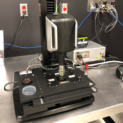
The RS-G4 has been designed with a compact, high-speed resonance scanner and scanning stage with macro and micro-imaging optics that allow the acquisition of in-vivo and ex-vivo images from slides to cleared whole tissue mounts in the micrometer to centimeter scales.
In addition, ex-vivo tissue sections can be imaged in both reflectance and fluorescence modes. Its 360-degree rotation of the scan head allows greater flexibility and precise angling for acquiring images of large tissue, organs, or whole animals. With a single nose piece, the instrument is essentially agnostic as to what type of objectives can be used. We routinely use our Nikon Plan Apo 10xC/0.5 NA (WD: 5.5 mm) and the Nikon CFI90 20xC/1.0 NA (WD: 8.2 mm) glycerol immersion objectives for imaging cleared tissue.
References
Equipment Specifications
We have objective options from 1x to 100x, including the Nikon CFI90 20xC/1.0 NA (WD: 8.2 mm) objective designed for cleared samples. Resolution is determined by which objective is used with individual frames (1024 x 1024 pixels) captured at up to 25fps.
Highlights
- Supports air, glycerol, water, and oil immersion objectives
- Fast, large-area mosaic creation up to 120 x 80 mm x 11mm
- Simultaneous and sequential imaging for reflectance and fluorescence
- Experiment acquisition setup for snapshots, mosaics, time, Z-series, and multi-points. wavelength
Lasers
- Wavelengths include: 405 nm, 488 nm, 561 nm, 638 nm, 785 nm
Objectives
|
Magnification |
NA |
Correction |
Optic Properties |
Tube Length/Cover Glass Thickness |
Working Distance (mm) |
Immersion Medium |
|---|---|---|---|---|---|---|
|
2x |
0.10 |
PlanApo λ |
|
8.5 |
Air |
|
|
4x |
0.20 |
PlanApo |
|
∞/- |
15.7 |
Air |
|
10x |
0.50 |
PlanApo |
Correction Collar |
∞/- |
5.5 |
Glycerol+ |
|
10x |
0.45 |
PlanApo |
DIC N1 |
∞/0.17 |
4 |
Air |
|
20x |
1.00 |
Correction Collar |
∞/0 |
8.2 |
Glycerol+ |
|
|
ELWD 20x |
0.45 |
S Plan Fluor |
Correction Collar/ Ph1 Adm |
∞/0-2 |
8.2-6.9 |
Air |
Leica SP5 Upright Confocal Microscope
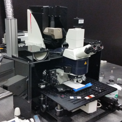
Leica DM6000 CFS upright multiphoton microscope equipped with a TCS SP5 MP laser confocal with both galvonomic and resonance scanners and with internal spectral HyD detectors. 405, 458, 476, 488, 514, 543, 594, and 640 nm single photon laser lines and a Mai Tai eHP DS 690-1040 nm tunable multiphoton laser along with 4-channel non-descanned detectors. Objectives available include 63x/NA 1.4 oil and 25x/NA 0.95 water (optimal for intravital deep tissue imaging (up to 2.5 mm)).
Objectives
|
Magnification |
NA |
Correction |
Optic Properties |
Tube Length/Cover Glass Thickness |
Working Distance (mm) |
Immersion Medium |
|---|---|---|---|---|---|---|
|
5x |
0.15 |
HCX PL FLUOTAR/CS |
|
∞/-/C |
13.7 |
Air |
|
10x |
0.4 |
HC PL APO/CS |
Confocal Series |
∞/0.17/A |
2.2 |
Air |
|
20x |
0.7 |
IMM CORR/CS |
Confocal Series |
∞/-/C |
0.26 |
Water-Glycerol-Oil |
|
L 25x |
0.95 |
HCX IR APO/CS |
VISIR |
∞/0.17/0 |
2.4 |
Water Immersion |
|
L 25x |
0.95 |
HC FLUOTAR |
VISIR |
∞/0/0 |
2.5 |
Water Dipping |
|
40x |
1.3 |
HCX PL APO/CS |
Confocal Series |
∞/0.17/0 |
0.24 |
Oil |
|
63x |
1.4-0.60 |
HCX PL APO/CS |
Confocal Series |
∞/0.17/E |
0.14 |
Oil |
Jackson Hall
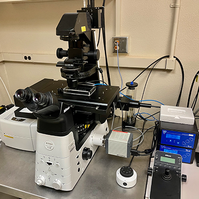
Nikon AX R
Nikon Ti2: Fully motorized (multi-position nosepiece, filters, condenser, z-motor, x/y stage) inverted microscope with 25 mm field of view. This FAST resonant (15 fps full 2K FOV with up to 720 fps at 2048x16 pixels) or galvanomic scanners (up to 8192x8192 pixels for Nyquist resolution at lower magnifications and the larger FOV). This scope has Nikon’s Perfect Focus System.
Live Cell Imaging: Ibidi stage top incubator and environmental controls enabling researchers to perform live cell imaging in plates, slides and dishes
LED light source excitation (in nm): 390, 440, 475, 510, 555, 575, 637, 748
Widefield Camera: Teledyne Photometrics Prime 95B sCMOS
Confocal Imaging lasers and detection filters for: 408, 488, 561 and 640 nm excitation lines
Widefield Emission Filters:
Widefield illumination and filters for a variety of fluorochromes including the Opal dyes UV, CFP, YFP, GFP, dsRed, TRITC, CY5 imaging
| Filter Name | Peak (nm) | Range (nm) |
| DAPI | 432 | 36 |
| Opal 520 | 515 | 30 |
| Opal 540 | 544 | 24 |
| Opal 570 | 595 | 31 |
| Opal 620 | 641 | 75 |
| Opal 690 | 680 | 42 |
Objectives:
| Magnification | NA | WD (mm) | Immersion Medium |
| 2x | .10 | 8.5 | Air |
| 4x | .1 | 16.5 | Air |
| 10x | .3 | 15.2 | Air |
| 20x | .7 | 2.3-1.3 | Air |
| 20x LWD* | .95 | 0.23-0.11 | Water |
| 40x | .95 | 0.99-0.9 | Air |
| 40x ELWD* | .6 | 3.6-2.8 | Air |
| 60x | 1.4 | 0.13 | Oil |
| 100x* | 1.49 | 0.13 | Oil |
| 100x* | 1.35 | 0.31-0.28 | Silicone |
| *(available by request) | |||
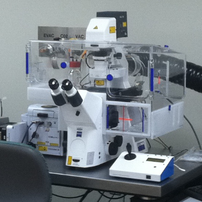
Nikon A1RHD Multiphoton Microscope
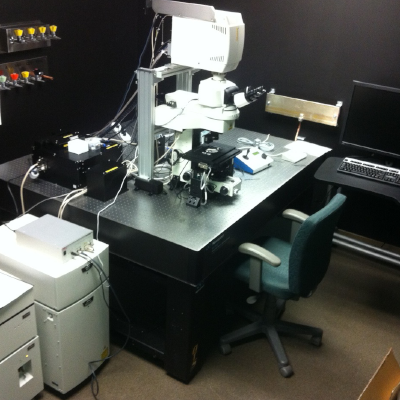
Nikon A1RMP confocal is connected to an upright Nikon FN1 microscope featuring a Plan Apo LWD 25x water immersion, NA 1.1 objective most suitable for imaging deep (it has a 2 mm working distance) into a fixed or living sample. In addition to laser scanning confocal imaging, conventional, and resonant visible laser confocal imaging, a Spectra-Physics 15W Mai Tai eHP tunable IR laser and six, three GaAsPs (epi direction) and three conventional PMTs (trans direction) non-descanned GaAsP detectors allow multiphoton visualization of multiple fluorophores located deep within the specimen. Visible lasers include 405, 457, 488, 514, 561 and 637 nm lines. The system has both galvonomic (4K) and resonant (1k, 8Khz, up to 720fps) scanners that can be used in concert simultaneously for photoactivation and MP imaging. A encoded Prior motorized stage and 200 µm Piezo objective motor can be used for tiling or multiple-position imaging.
The system is also equipped with Second Harmonic Generation (SHG) detection. SHG doesn't involve the excitation of molecules like other techniques such as fluorescence microscopy. See UIC staff for training and questions.
This instrument was funded in 2012 by the Minnesota Partnership in conjunction with the Mayo Clinic Foundation, Rochester, MN.
In October 2018 the system was upgraded to an A1RHD with 1024 resonant scanning and improved signal-to-noise imaging in resonant mode.
Objectives
|
Magnification |
NA |
Correction |
Optic Properties |
Tube Length/Cover Glass Thickness |
Working Distance (mm) |
Immersion |
|---|---|---|---|---|---|---|
|
10x |
0.3 |
PlanFluor |
DIC L |
∞/0.17 |
16 |
Dry |
|
10x |
0.3 |
Fluor |
DIC L |
∞/0 |
2 |
Water |
|
10x |
0.5 |
Plan Apo |
Correction Collar |
∞/- |
5.5 |
Glycerol |
|
20x |
0.45 |
PlanFluor |
DIC L |
∞/0-2 |
7.4 |
Dry |
|
20x |
0.75 |
PlanApo lambda |
DIC N2 |
∞/0.17 |
1 |
Dry |
|
25x |
1.1 |
ApoLWD |
DIC 2, Correction Collar |
∞/0-0.17 |
2 |
Water |
|
40x |
0.8 |
Fluor |
DIC M |
∞/0 |
2 |
Water |
|
100x |
1.3 |
PlanFluor |
|
∞/0.17 |
0.2 |
Oil |
Filters - MP
| Filter cube | Emission band | Common dyes |
|---|---|---|
| 99020 | 450/40 | DAPI |
| 9920-IR | 460/50 | DAPI |
| 99022 | 525/50 | GFP, AF488 |
| 99021 | 485/30 | CFP |
| 99023 | 600/50 | mCherry, AF568 |
| 99023 | 660LP | AF647, Cy5 |
| 99024 | 545/40 | YFP |
Filters - MP NDD
| Filter cube | Emission band | Common dyes |
|---|---|---|
| Epi NDDs | ||
| 99124 | 425/50 |
SHG collagen, DAPI TPE |
| 99121 | 510/80 | GFP |
| 99121 | 605/70 | mCherry |
| Trasn NDDs | ||
| 99020 | 450/40 |
SHG collagen, DAPI TPE |
| 99121 | 515/70 | GFP |
| 99121 | 605/70 | mCherry |
| Available on demand | ||
| 99321 | 440/80 |
SHG collagen, DAPI TPE |
| 99324 | 540/80 | YFP |
| 99324 | 620/52 | mCherry |
Nikon A1Rsi HD Confocal with SIM Super Resolution
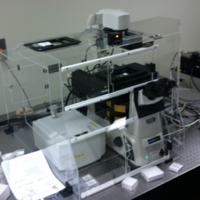
Nikon Ti-E Motorized Microscope featuring PFS Perfect Focus III focus lock technology. LED Illumination systems for both transmitted and fluorescence light applications. Objectives at 10x, 20x, 40x, 60x (water and oil immersion), and 100x magnifications for DIC. Fluorescence accessories for widefield applications for DAPI, GFP, RFP, and Cy5. The Nikon Elements software has the JOBS package that can automate image collection for high-content screening of plates.
SIM Structured Illumination System-Using high-frequency Structured Illumination, the Nikon N-SIM can achieve an image resolution of 120 nm, which was previously considered impossible with optical microscopes. Furthermore, with a temporal resolution of up to 0.6 sec/frame, N-SIM enables super-resolution time-lapse imaging capture of dynamic molecular interactions in living cells.
- Lasers (A1R): 405 nm, 488 nm, 561 nm, 640 nm
- Lasers (N-SIM): 405 nm, 488, nm, 561 nm, 640 nm
- Detectors: The A1R has four PMTs for confocal detection, two of them GaAsPs, with double the sensitivity of traditional PMTs. There is also a transmitted light detector and a 32-channel spectral detector.
- N-SIM: Hamamatsu FLASH 4
Objectives
|
Magnification |
NA |
Correction |
Optic Properties |
Tube Length/Cover Glass Thickness |
Working Distance (mm) |
Immersion |
|---|---|---|---|---|---|---|
|
2x |
0.1 |
PlanApo |
8.5 mm |
Dry |
||
|
10x |
0.45 |
PlanApo |
DIC N1 |
∞/0.17 |
4 |
Dry |
|
20x |
0.75 |
PlanApo VC |
DIC N2 |
∞/0.17 |
1 |
Dry |
|
40x |
0.6 |
S PlanFluor |
DIC N1, Correction Collar |
∞/0-2 |
3.6 |
Dry |
|
40x |
0.95 |
PlanApo |
DIC MN 2, Correction collar |
∞/0.11-0.23 |
0.14 |
Dry |
|
60x |
1.27 |
PlanApo IR with correction collar |
DIC N2 |
∞/0.15-0.19 |
0.18-0.16 |
Water |
|
60x |
1.4 |
Plan Apo Lambda |
DIC N2 |
∞/0.17 |
0.13 |
Oil |
|
100x |
1.49 |
SR ApoTIRF with correction collar |
DIC N2 |
∞/0.13 |
0.12 |
Oil |
Filters - Confocal
| Filter cube | Emission band | Common dyes |
|---|---|---|
| 99020 | 450/40 | DAPI |
| 99022 | 525/50 | GFP, AF488 |
| 99023 | 600/50 | mCherry, AF568 |
| 99023 | 660LP | AF647, Cy5 |
Filters - Camera
| Filter cube | Excitation band | Dichromatic mirror | Emission band | Common dyes |
|---|---|---|---|---|
| Installed | ||||
| Chroma 49028 | 395/25 | T425lprx | 460/50 | DAPI |
| Chroma 49002 | 470/40 | T495lprx | 525/50 | GFP, AF488 |
| Chroma 49005 | 545/30 | T570lp | 620/60 | dsRed, AF568 |
| Chroma 49006 | 620/60 | T660lprx | 700/75 | AF647, Cy5 |
| Available on demand | ||||
| Chroma 49303 | 495/25 | T515lprx | 537/29 | Opal 520 |
| Chroma 49304 | 546/10 | T556lprx | 572/23 | Opal 570 |
| Chroma 49306 | 580/25 | T600lprx | 615/30 | Opal 620 |
| Opal 620 | 590/20 | 635/75 | Opal 620 | |
| Chroma 49022 | 650/45 | T685lprx | 720/60 | Opal 690, Cy5.5 |
Filters - SIM
| Filter cube | Excitation band | Dichromatic mirror | Emission band | Effective emission window | Common dyes |
|---|---|---|---|---|---|
| Installed | |||||
| SIM405 | 395/20 | 440LP | 460/50 | 440-485 | DAPI |
| SIM488 | 480/20 | 515LP | 523/45 | 515-545 | GFP, AF488 |
| SIM561 | 561/10 | 590LP | 605/70 | 590-640 | mCherry, AF568 |
| SIM640 | 620/60 | 665LP | 700/75 | 665-738 | AF647, Cy5 |
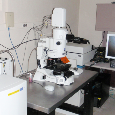
Olympus FLUOVIEW FV1000 BX2 Upright Confocal
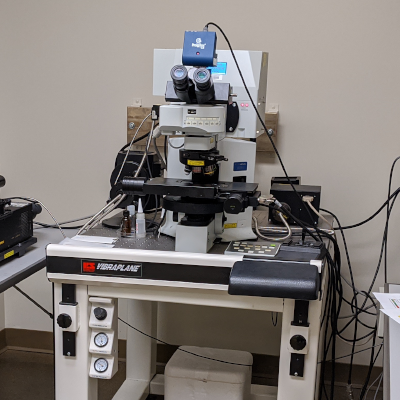
Olympus FluoView FV1000 is designed for high-resolution, confocal observation of both fixed and living cells. The FV1000 offers advances in confocal system performance while providing the speed and sensitivity required for live cell imaging, with minimal risk of damage to living specimens. The system has four fluorescence detectors plus a transmitted light detector for DIC.
- Automated upright BX61 microscope base
- 100W Mercury fluorescent lamp
- 100W halogen transmitted illuminator with detector
- PRIOR ProScanII motorized XY stage
- DIC optics
Methods
- Fluorescence, Brightfield, DIC, FRAP, FRET
Automated Capabilities
- Time-lapse, Multi-channel fluorescence, Co-localization, Z-stacks
Lasers
- 405nm 25mW Diode, 458, 488, 515nm 30mW Argon, 543nm 1mW HeNe green, 635nm 10mW HeNe red
Detector
- 4 photomultiplier tubes channels, 1 transmission detector
Objectives
|
Magnification |
NA |
Correction |
Optic Properties |
Tube Length/Cover Glass Thickness |
Working Distance (mm) |
Immersion |
|---|---|---|---|---|---|---|
|
4x |
0.16 |
UPlanApo |
-- |
∞/0.17 |
13 |
Dry |
|
10x |
0.4 |
UPlanApo |
BX2-DIC10 |
∞/0.17 |
3.1 |
Dry |
|
20x |
0.85 |
UPlanSApo |
BX2-DIC20 |
∞/-/FN26.5 |
0.17 |
Oil |
|
40x |
1.3 |
UPlanFLN |
BX2-DIC40 |
∞/0.17/FN26.5 |
0.2 |
Oil |
|
60x |
1.42 |
UPlanApo N |
BX2-DIC60 |
∞/0.17/FN26.5 |
0.15 |
Oil |
|
60x |
0.9 |
LUMPlanFL |
|
∞/0 |
2 |
Water |
Filters
|
Filter |
Excitation Filter (nm) |
Dichroic Mirror (nm) |
Emission Filter (nm) |
|---|---|---|---|
|
DAPI |
D350/50 |
BS20/80 |
460/50 |
|
FITC |
D480/30 |
DM405/488 |
D535/40 |
|
TRITC |
D540/25 |
DM405/488/543/635 |
D605/55 |
|
Cy5 |
D620/20 |
DM458/515 |
D700/75 |
Channels
|
Channels |
Dichroic Mirror (nm) |
Barrier Filters (nm) |
|---|---|---|
|
Channel 1 |
SDM560, SDM510, SDM490 |
BA505-525, BA480-495, BA430-470 |
|
Channel 2 |
SDM560 |
BA505-605, BA560-620, BA535-565, BA505-525 |
|
Channel 3 |
SDM640 |
BA560-660 |
|
Channel 4 |
-- |
BA655-755 |
Zeiss Cell Observer Spinning Disk Confocal
Zeiss Cell Observer Spinning Disk Confocal
The Zeiss Cell Observer Spinning Disk confocal fluorescence microscope for imaging live samples at fast speed. Definite focus, time-lapse, multi-well, and tiling of images. DIC and phase contrast capable. Full environmental chamber for temperature and atmosphere control. Three laser lines are available (405, 488, and 561 nm).
Motorized Zeiss Axio Observer Z1 inverted scope with temperature-controlled enclosure, CO2, Definite Focus, multi-channel acquisition, piezo x/y/z stage.
Methods
- Fluorescence, Brightfield, Phase contrast, DIC, Time Lapse, Live Cell with 37°C / CO2 enclosure
Automated Capabilities
- Time-lapse, Stitching, Multi-channel fluorescence, Co-localization, Piezo Z-stacks
Lasers
- 405 nm, 50 mW, 488 nm, 100 mW, 561 nm, 30 mW
Detector
- Photometrics QuantEM 512SC - High Sensitivity 16 bit, 512 x 512 grayscale CCD
Objectives
|
Magnification |
NA |
Correction |
Optic Properties |
Tube Length/Cover Glass Thickness |
Working Distance (mm) |
Immersion Medium |
|---|---|---|---|---|---|---|
|
10x |
0.3 |
EC Plan-NEOFLUAR |
Ph1 |
∞/- |
3.2 |
Air |
|
20x |
0.75 |
Plan-ApoCHROMAT |
Phase 2, DIC II |
∞/0.17 |
0.61 |
Air |
|
40x |
0.95 |
EC Plan-APOCHROMAT |
KORR |
∞/0.13-0.21 |
|
Air |
|
63x |
1.2 |
C-ApoCHROMAT |
KORR UV-VIS-IR, Correction Collar |
∞/0.17 |
0.28 |
Water Immersion |
|
100x |
1.4 |
Plan-ApoCHROMAT |
DIC |
∞/0.17 |
0.11 |
Oil Immersion |
Snyder Hall
Nikon AX R
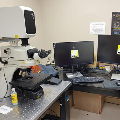
- Nikon NiE fully motorized (multi-position nosepiece, filters, condenser, z-motor, x/y stage) upright microscope with 25 mm field of view. This resonant (15 fps full FOV with up to 720 fps at 2048x16 pixels) and galvanomic scanners (up to 8192x8192 pixels for Nyquist resolution at lower magnifications or larger FOVs)
- Lasers and detection filters utilizing the 408, 445, 488, 514, 561, and 640 nm excitation lines allow for multiplexed imaging of DAPI, CFP, YFP, GFP, FITC, dsRed, TRITC, CY5, and many more fluorescent reporters.
- Four-channel GaAsP PMT detector modules, including spectral detection along with transmitted light.
- Objectives 2x, 4x, 10x, 20x, 40x, 60x, 100x including water, silicon and oil immersion options. The system is compatible with many of the UIC’s extensive collection of additional Nikon objectives.
- Differential Interference Contrast (DIC)
- Nikon Element ER software has built-in multidimensional (multi-XY, Z, T, multichannel) experiment capabilities. The JOBS module allows customization in setting up non-orthogonal experiments with multiple paths and dimensions. Analysis of data can be done in real-time during the experiment, and the direction of the experiment can even be changed based on the results of the analysis. Fully automated collection and analysis capabilities with JOBS. The UIC staff is excited to help train you from the most basic image acquisition to the full complement of our instrument's capabilities.
Nikon A1si Confocal and Widefield Microscope
The Nikon A1si is a confocal system equipped with a point-scan confocal scanner, four PMT detectors for fluorescence and one for transmitted light. The scope also has a 32-channel PMT spectral detector. The A1si's highest pixel resolution mode is up to 4096 x 4096 pixels. The system is mounted on a Nikon Eclipse Ti2 inverted scope equipped with Perfect Focus System, fluorescence filters with DIC optics and a widefield Hamamatsu Flash 4 camera. There is also an IBIDI environmental stage top incubator system for long-term live cell imaging. Nikon NIS Elements imaging software is used to control acquisition and analysis. Nikon JOBS will permit investigators to create complex automated image acquisitions with intelligent analysis.
The advantages of multispectral imaging and unmixing have been shown in hundreds of publications to reduce autofluorescence by as much as 100-fold, both for microscopy and FFPE tissues and in small animal and plant fluorescence imaging.
Lasers
- 405 nm diode at 38mW, Argon multiline laser 457/477/488/514nm at 40mW, 561 nm diode at 40mW, 638 nm diode at 10mW
Detectors
- 4 PMTs, a Transmitted light detector, and a 32-channel spectral detector
References
Tam, et al "Improved in vivo whole-animal detection limits of green fluorescent protein-expressing tumor lines by spectral fluorescence imaging" Molecular Imaging, 6, 2007, 269.
Mansfield, et al "Visualization of microscopy-based spectral imaging data from multi-label tissue sections" Current Protocols in Molecular Biology, 2008 Oct; Chapter 14:Unit 14.19. DOI: 10.1002/0471142727.mb1419s84.
Levenson, et al "Multispectral imaging in biology and medicine: slices of life." Cytometry A. 2006 Aug 1;69(8):748-58.
Objectives
|
Magnification |
NA |
Correction |
Optic Properties |
Tube Length/Cover Glass Thickness |
Working Distance (mm) |
Immersion Medium |
|---|---|---|---|---|---|---|
|
4x |
0.2 |
Plan Apo |
-- |
∞/-- |
20 |
Air |
|
10x |
0.3 |
Plan Apo |
-- |
∞/0.17 |
4 |
Air |
|
20x |
0.75 |
Plan Apo |
DIC N2 |
∞/0.17 |
1 |
Air |
|
60x |
1.2 |
Plan Apo |
DIC N2, Correction collar |
∞/0.15-0.18 |
0.27 |
Water |
|
60x |
1.4 |
Plan Apo |
DIC H |
∞/0.17 |
0.21 |
Oil |
Filters
Widefield Filters
|
Common Fluorophore |
Excitation Filter (nm) | Dichroic Mirror (nm) | Emission Filter (nm) |
|---|---|---|---|
|
DAPI |
340-380 | 400LP | 435-485 |
|
CFP |
426-446 | 455LP | 460-500 |
|
GFP |
465-495 | 505LP | 515-555 |
|
YFP |
490-515 | 520LP | 520-560 |
| Texas Red | 532-587 | 595LP | 608-683 |
Confocal Filters
|
Common Fluorophore |
Excitation Filter (nm) | Dichroic Mirror (nm) | Emission Filter (nm) |
|---|---|---|---|
|
DAPI |
DS350/50 | BS20/80 | 460/50 |
|
CFP |
DS440/40 | 400-457/514 | 470/40 |
|
GFP |
D480/30 | DM405/488 | D535/40 |
|
YFP |
DS 510/50 | 400-457/514 | D 555/30 |
| dsRed | DS540/25 | 408/488/543 | D600/50 |
| Cy5 | DS640/20 | DM458/515 | D685/70 |
Nikon AZ100 C1si Spectral Confocal Microscope
The AZ100 Multizoom Macroscope with the C1si Spectral Confocal attachment makes the ultimate platform for in vivo imaging. Ideal for plant biology, developmental biology, cell biology, stem cell, and tissue research, the AZ-C1si allows the researcher to view large specimens in confocal mode. The AZ-C1si can capture fields of view up to 1cm and permits deeper confocal imaging than conventional microscopes thanks to its large working distance objectives. Whole organisms can be monitored and documented over time (for example, embryos) offering a wealth of continuous information on development or the organism’s response to experimental variables. Both conventional PMTs and Spectral detectors are installed on this instrument.
The system is equipped with 1x, 4x, and 5x objectives with variable optical zoom. There are 405, 457, 476, 488, 514, 561, and 637 nm lasers for image, co-localization, FRAP, FRET, and spectral studies.
The system is run using Nikon Elements Software.
The advantages of multispectral imaging and unmixing have been shown in hundreds of publications to reduce autofluorescence by as much as 100-fold, both for microscopy and FFPE tissues and in small animal and plant fluorescence imaging.
Lasers
405 nm, 457 nm, 488 nm, 514 nm, 567 nm, 637 nm
References
Tam, et al "Improved in vivo whole-animal detection limits of green fluorescent protein-expressing tumor lines by spectral fluorescence imaging" Molecular Imaging, 6, 2007, 269.
Mansfield, et al "Visualization of microscopy-based spectral imaging data from multi-label tissue sections" Current Protocols in Molecular Biology, 2008 Oct; Chapter 14:Unit 14.19. DOI: 10.1002/0471142727.mb1419s84.
Levenson, et al "Multispectral imaging in biology and medicine: slices of life." Cytometry A. 2006 Aug 1;69(8):748-58.
Objectives
|
Magnification |
NA |
Correction |
Optic Properties |
Tube Length/Cover Glass Thickness |
Working Distance (mm) |
Immersion Medium |
|---|---|---|---|---|---|---|
|
1x |
0.1 |
AZ Plan Apo |
|
|
35 mm |
Air |
|
4x |
0.4 |
AZ Plan Apo |
|
|
20 mm |
Air |
|
5x |
0.5 |
AZ Plan Fluor |
|
|
15 mm |
Air |
Filters
|
Filter |
Wavelengths |
||
|---|---|---|---|
|
Excitation Filter (nm) |
Dichoric Mirror (nm) |
Emission Filter (nm) |
|
|
DAPI |
D350/50 |
BS20/80 |
460/50 |
|
GFP |
D480/30 |
DM405/488 |
D535/40 |
|
TRITC |
D540/25 |
405/488/543 |
D605/55 |
|
Cy5 |
D640/20 |
DM458/515 |
D680/30 |
Channels
|
Channels |
Dichoric Mirror (nm) |
Barrier Filters (nm) |
|---|---|---|
|
Channel 1 |
SDM560, SDM510, SDM490 |
2nm Spectral Monchrometer Grating |
|
Channel 2 |
SDM640, SDM560 |
2nm Spectral Monchrometer Grating |
|
Channel 3 |
SDM640 |
BA560-620, BA560-660, BA655-755 |
Wallin Medical Biosciences Building (WMBB)
Leica Stellaris 8
Leica DMi8 inverted laser scanning confocal microscope with both galvonomic and resonance scanners and 5 spectral internal HyD-S detectors. A fixed 405nm violet laser and pulsed white-light laser (WLL) that is tunable from 440-790nm allows for precise excitation control and multi-spectral imaging. The WLL and HyD-S detectors allow for Leica's patented Tau gating and separation modalities using fluorescence lifetime measurements. Environmental control enables long-term live cell imaging and the Leica Navigator interface streamlines stage control and multi-position high-speed tiling.
Objectives include 20x/0.7NA multi-immersion, 40x 1.25NA glycerol motCORR, 63x 1.4NA oil, and 63x 1.2NA water.