Faculty


Bio
Expertise
- Arbovirus virology
- Vector biology
- Animal model development
- Emerging infectious diseases
- Host-pathogen interactions
Professional Associations
- Forward Under 40 Award, UW Alumni Association
- NIH Postdoctoral Fellowship in Biodefense and Emerging Infectious Disease
- Richard F. Marsh Outstanding Graduate Student Award
UMN Professional Associations
- Microbiology, Immunology, and Cancer Biology (MICaB) Ph.D. program
- Institute for Molecular Virology
Research Summary
Research summary/interests
Many arthropod-borne viruses (arboviruses) are resurgent, are spreading to new environments, and are responsible for substantial morbidity and mortality around the globe as climate change and urbanization enhance opportunities for spread and interspecies transmission. My primary research goals are to understand commonalities in patterns of arbovirus population evolution, how these features account for their success in emergence and epidemics, and how these evolutionary processes can be exploited to prevent disease. The underlying factors that promote (re)emergence are poorly understood; therefore, my team utilizes basic and applied research that emphasizes understanding the molecular and cellular mechanisms involved in establishing and maintaining the host-pathogen relationship, the role of host-specific and pathogen-specific evolutionary pressures in defining these relationships, and understanding the mechanisms of inter-species transmission. These areas primarily focus on Zika virus; however, four other arboviruses transmitted by Aedes aegypti- dengue, yellow fever, Mayaro, and chikungunya- remain circulating in the Americas, and my research interests align with all of these viruses. If the current Zika virus situation has taught us anything, it is that arbovirus emergence/re-emergence is the “new normal” in the Americas. Future outbreaks are unpredictable but likely are inevitable. Therefore, our overall goal is to develop strategies to interrupt transmission or predict a zoonotic pathogen’s adaptability or evolvability.
Research funding grants
- NIH/NIAID Exploratory/Development Research Grant R21
- NIH/NIAD Research Project Grant R01
- NIH/NIAID Program Project Grant P01
- NIH/NIAID High Priority, Short-term Project Award R56
Teaching Summary
Mentoring statement
I am a strong believer that the best science comes from a happy learning environment, and that we will use fundamental biological principles to think critically about challenges facing our society and the world.
Academic interests and focus
- Vector biology
- Ecology and evolution of infectious diseases
- Emerging infectious diseases
Teaching areas
DVM, graduate, and undergraduate courses
Courses
CFAN 3334, Parasites and Pestilence

Bio
Bryce A. Binstadt, MD, PhD, is an Professor of Pediatrics in the Division of Pediatric Rheumatology, Allergy, and Immunology and a Distinguished University Teaching Professor. Dr. Binstadt cares for children with arthritis, systemic lupus erythematosus, dermatomyositis, and related rheumatologic disorders. He heads a laboratory in the University of Minnesota's Center for Immunology focused on understanding the pathogenesis of autoimmune diseases with particular emphasis on immune-mediated cardiovascular disease. He is the Director of the Pediatric Rheumatology Fellowship Training Program, an Associate Director of the Medical Scientist Training Program (MD/PhD), and Director of the Pediatric Physician Scientist Training Program.
Dr. Binstadt received his MD degree from Mayo Medical School and his PhD in Immunology from Mayo Graduate School in Rochester, MN. He then completed his residency in the Boston Combined Residency Program in Pediatrics at Boston Children's Hospital and Boston Medical Center, followed by fellowship training in Pediatric Rheumatology at Boston Children's Hospital. He served as an attending physician in the Rheumatology Program at Boston Children's Hospital/Harvard Medical School as well as a research fellow in the Section on Immunology and Immunogenetics at the Joslin Diabetes Center for two years before joining the faculty at the University of Minnesota in 2007. Dr. Binstadt is board-certified in Pediatrics and Pediatric Rheumatology.
Research Summary
Autoimmunity, Innate Immunity
The Binstadt Laboratory is broadly interested in autoimmune and rheumatic disease pathogenesis.
The main area of investigation focuses on how systemic autoimmune diseases lead to cardiovascular inflammation and damage. We use the K/BxN T cell receptor (TCR) transgenic mouse model. K/BxN mice spontaneously develop both inflammatory arthritis and autoimmune valvular carditis, similar to patients with rheumatic heart disease or systemic lupus erythematosus. Current projects seek to understand how macrophages, fibroblasts, and endothelial cells promote progression of cardiac valve inflammation and fibrosis.
A second project focuses on developing and testing novel inhibitors of the arthritis-promoting cytokine TNF.
A new collaborative project explores cardiovascular inflammation that occurs in lysosomal storage diseases, particularly mucopolysaccharidosis type I (MPS-I).
Current Funding Sources:
- National Institutes of Health (NIH)
- American College of Rheumatology/Rheumatology Research Foundation
Clinical Summary
Juvenile dermatomyositis; Juvenile rheumatoid arthritis; Pediatric autoimmune diseases; Systemic lupus erythematosus
Education
Fellowships, Residencies, and Visiting Engagements
Licensures and Certifications
Honors and Recognition
Contact
Address
Pediatric Rheumatology, Allergy, & ImmunologyAcademic Office Building
2450 Riverside Ave S AO-10
Minneapolis, MN 55454

Bio
Dr. Blazar is a Regents Professor of Pediatrics in the Division of Blood and Marrow Transplant & Cellular Therapy and attends on the Pediatric Blood and Marrow Transplantation (BMT) service. Dr. Blazar is the recipient of the Children’s Cancer Research Fund Land Grant Chair in Pediatric Oncology to recognize his pioneering work in the development of novel immune-based therapies. He is the Founding Director of the Clinical and Translational Sciences Institute and the Founding Director of the Center for Translational Medicine.
Dr. Blazar received his MD from Albany Medical College. He completed a residency in Pediatrics and a fellowship in hematology/oncology and bone marrow transplantation at the University of Minnesota. Dr. Blazar joined the University of Minnesota faculty in 1985. He is board certified in Pediatrics and Hematology/Oncology.
Dr. Blazar is the recipient of National Institutes of Health (NIH) MERIT Awards from the National Heart, Lung, and Blood Institute as well as the National Institute of Allergy and Infectious Diseases. He is the Principal Investigator of the UMN Clinical and Translational Sciences Award (U54), NIH funded R01 grants, P01 Projects, a U19 grant subcontract and Leukemia and Lymphoma Translational Research grants focusing on BMT immunological studies. Dr. Blazar is the author of more than 725 manuscripts, which have appeared in premier peer-reviewed publications.
Research Summary
1. Graft-versus-host disease (GVHD) . GVHD is a multi-organ system disorder in which donor T cells recognize host alloantigens present on antigen-presenting cells and tissues in the context of an inflammatory response. Studies are directed toward identifying and modifying signals that drive or inhibit acute and chronic GVHD generation. These include the analysis of positive costimulatory molecules and negative regulators of the immune response that counterbalance positive costimulation as well as intracellular signaling and metabolic pathways that regulate these responses at the level of the GVHD target organ. We are examining cell-based therapies such as regulatory T cells (see below) in mice and in patients and myeloid-derived suppressor cells (see below). We have also used a newly developed model of chronic GVHD that results from T:B cooperativity, leading to alloantibody and subsequently, collagen deposition, culminating in multi-organ system injury and pulmonary and liver fibrosis. We have tested drugs and cell populations that affect this interaction, many of which have been brought into the clinic, one of which has received FDA approval. For approaches that reduce acute or chronic GVHD in mice, we are assessing their immune competency to malignant cells (to assess graft-versus-leukemia, GVL) and to pathogens.
2. Regulatory cells. A) regulatory T cells (Treg) . Using phosphoproteomics, metabolomics, micro-RNA/mRNA binding partners and flow cytometry, we have identified pathways that can be targeted to upregulate Treg potency in mice and humans. Proof-of-principle has been demonstrated in allogeneic and xenogeneic mouse models . Other studies are focused on genome engineering approaches that should optimize GVHD and autoimmune disease treatment. Knockout, transgenic and conditional inducible strains of mice are utilized along with 2-photon microscopy to identify mechanisms of in vivo suppression. B) Myeloid-derived suppressor cells (MDSC). We have developed techniques to differentiation and active MDSCs from normal hematopoietic sources in mice and humans. Our studies have shown in murine allogeneic GVHD systems that inflammasome activation converts MDSCs into immune stimulatory cells. Knockout of inflammasome components or inflammasome inducers have ameliorated this loss of function. Ongoing studies are examining the production of human MDSCs from inducible pluriopotent stem cells, with or without genome engineering approaches, for mechanistic purposes and potential future clinical trials.
3. T cell generation from induced pluripotent stem cells (IPSCs). In order to understand the optimal requirements for T cell anti-cancer or anti-pathogen responses, we have focused on reprogramming various human cell populations into thymic progenitor cells and mature T cells. Transcriptomics and epigenetics are investigated. Genome modification of IPSCs readily induces chimeric antigen receptor (CAR) expression that directs T cells to lymphoma cells. Using a model that incorporates a CAR used in the clinic for treating B cell malignancies and a transgenic strain that expresses human B cell antigens, cytokine release syndrome and neurotoxicity have been shown. Studies are being examined to explore different approaches to mitigate these side-effects.
Clinical Summary
Pediatric hematology/oncology
Bone marrow transplant
Graft-versus-host disease
Graft-versus-leukemia
Tumor immunotherapy
Education
Fellowships, Residencies, and Visiting Engagements
Licensures and Certifications
Honors and Recognition
Contact
Address
Pediatric Blood and Marrow Transplantation & Cellular TherapyMayo Mail Code 366
420 Delaware Street SE
Minneapolis, MN 55455
Administrative Contact
Colette Brosko
Administrative Phone: 612-624-7920
Administrative Email: cbrosko@umn.edu
Administrative Fax Number: 612-624-3913


Bio
Administrator Info
Phone: 612-625-6911
Fax: 612-625-4410
Email: tbold@umn.edu
Mail: Wallin Medical Biosciences Building, MMC 2641, 2101 6th Street S.E., Minneapolis, MN 55414
Summary
Dr. Bold is a physician-scientist, trained in the clinical subspecialty practice of infectious diseases, and laboratory based investigations of microbiology and immunology. His clinical interests regard the care of immune-compromised patients being treated for cancer or undergoing transplantation. His lab in the Center for Immunology is focused on advancing understanding of how the adaptive immune system combats infection with Mycobacterium tuberculosis, the bacterial pathogen responsible for human TB.
Research Summary
Dr. Bold is a physician-scientist, trained in the clinical practice of infectious diseases and laboratory based investigations of microbiology and immunology. His clinical interests regard the care of immune-compromised patients being treated for cancer or undergoing transplantation. His lab in the Center for Immunology is focused on advancing understanding of how the immune system combats infection with Mycobacterium tuberculosis, the bacterial pathogen responsible for human TB, and SARS-CoV-2, the virus that causes COVID-19.
Clinical Summary
Transplant Infectious Diseases
Education
Fellowships, Residencies, and Visiting Engagements


Bio
Administrator Info
Name: Admin
Phone: 612-624-9996
Fax: 612-625-4410
Email: idimdivision@umn.edu
Mail: Microbiology Research Building, 1st floor mail room, MMC 2821, 689 23rd Ave S.E., Minneapolis, MN 55455
Summary
Dr. Boulware is an infectious disease physician-scientist with formal training in clinical trials, public health, and tropical medicine. Dr. Boulware combines his clinical research with nested basic science investigations into disease pathogenesis to conduct translational research. His primary research interests are in meningitis in resource-limited areas including diagnosis, prevention, treatment, and quality improvement initiatives incorporating cost-effectiveness analyses in order to translate knowledge into improved care. Dr. Boulware's current research is focused on improving the clinical outcomes of HIV-infected persons with cryptococcal meningitis, the second most common AIDS-defining opportunistic infection in Sub-Saharan Africa and the most common cause of adult meningitis. Additionally, Dr. Boulware has been motivated to improve the diagnostics for TB meningitis, and now that TB meningitis can be promptly diagnosed, to also improve the treatment of tuberculous meningitis. Dr. Boulware leads a multidisciplinary, international research team with active research collaborations with partners in Uganda, South Africa, Ethiopia, Brazil, Botswana, the Netherlands, and the United Kingdom.
Research Summary
Reducing HIV-related mortality in people living with AIDS Improving Diagnostics for Meningitis Improving treatments for Cryptococcal meningitis and TB meningitis Preventing avoidable deaths due to subclinical cryptococcosis Clinical Trials for novel meningitis therapeutics and strategies Quality improvement initiatives to improve survival in resource-limited settings Antimicrobial resistance in low and middle income countries Research Projects: Improving Diagnostics and Neurocognitive Outcomes in HIV/AIDS-related Meningitis (R01 NS086312) Phased Implementation of a Public Health Programme: Cryptococcal Screening and Treatment in South Africa. ( R01AI118511) Operational Research for Cryptococcal Antigen Screening (ORCAS) of HIV Patients (U01AI125003) Infectious Disease Training in Clinical Investigation (T32AI055433) Encochleated Oral Amphotericin for Cryptococcal Meningitis Trial TB Meningitis: Evaluating CSF Immunology to Discover Hidden Disease and Potential Immunomodulatory Therapies Redefining Tuberculosis Meningitis with Metagenomics and Host Transcriptomics NIH ACTIV-6 COVID Platform Trial
Clinical Summary
Central nervous system infections; AIDS-related opportunistic infections; Tropical medicine; Travel medicine; Cost Effectiveness
Education
Fellowships, Residencies, and Visiting Engagements
Selected Publications
Research Summary
The goal of the Burrack lab is to develop a comprehensive understanding of host-pathogen interactions in order to enhance immunity and limit immune-mediated pathology with an emphasis on investigating the pathogenesis of malaria and other pathogens of global health importance. Malaria remains a major public health problem despite substantial efforts to reduce its associated morbidity and mortality. There are still around 200 million cases and over 400,000 deaths annually, with young children and pregnant women in sub-Saharan Africa experiencing the greatest burden. Our laboratory seeks to improve our knowledge of the immune response to Plasmodium infection and vaccination, which will inform the development of host-targeted therapeutics and vaccines to promote an effective immune response. We combine translational research and animal models of Plasmodium infection and vaccination to investigate the immune response to malaria. We investigate interactions between innate and adaptive immune cells during Plasmodium infection, with a focus on natural killer (NK) cells, T cells, and B cells. This is a highly tractable system to investigate the pathogenesis of Plasmodium infection and vaccination and test immunomodulatory therapeutics against this deadly disease.
Research Summary
Age is associated with increased inflammation, visceral adiposity and metabolic disease. Tissue resident immune cells are required for dampening inflammation and maintaining tissue homeostasis. There are changes in resident immune cells that drive the increased inflammation and metabolic impairments that are seen with increased age. We are studying the cellular and molecular changes within tissue resident immune cells that drive metabolic impairments in tissues. In particular, we are focused on lipolysis, a metabolic process that is required for release of energetic substrates from stored triglycerides in adipocytes. Lipolysis is impaired in aged individuals and this impairment may contributes to a worsened ability of elderly to maintain a healthy body-weight, stay warm or exercise. Our work has previously shown that adipose tissue immune cells reside in microenvironment niches and are able to inhibit lipolysis in the aged adipose tissue. There are two broad projects within the lab: Adipose tissue macrophage-specific regulatory effects on lipolysis and inflammation during aging Fat-associated lymphoid cluster (FALC) and lymphocyte regulation of metabolism Our lab focuses on mouse models of aging and uses a wide variety of techniques to investigate the changes occurring with age. We combine this in vivo approach with a complementary in vitro cell culture system to better understand a direct mechanism. Ultimately, our goal is to generate candidates that could be targets for therapeutically treating to improve health span and restore metabolism in the elderly.
Contact
Address
4-108 NHH312 Church St
Minneapolis, MN 55455

Bio
Administrator Info
Name: Ashley Fuchs
Phone: 612-624-0999
Email: fuchs@umn.edu
Fax: 612-625-2174
Summary
Monica Campo Patiño, MD, MPH., completed medical school at Universidad El Bosque in Bogotá, Colombia. She went on to complete a research fellowship at the Brigham and Women’s Hospital at Harvard Medical School in Boston, MA. She received a Master’s in Public Health at the Harvard T.H. Chan School of Public Health. Subsequently, she completed an internal medicine residency at Tufts Medical Center in Boston, MA and a fellowship in pulmonary and critical care medicine at the University of Washington in Seattle, WA.
Currently, Dr. Campo is an Assistant Professor in the University of Minnesota Department of Medicine in the Pulmonary, Critical Care, Allergy, and Sleep Medicine Division. Dr. Campo specializes in the treatment and management of general pulmonary and critically ill patients. She is committed to excellent and compassionate care of patients with chest problems. Dr. Campo’s research focuses on the discovery of new therapies for patients with respiratory infections. She has received independent funding from the National Institutes of Health for her research projects and has published in peer-reviewed journals as well as presented her research at international scientific meetings.
Research Summary
Dr. Campo’s research program focuses on the examination of regulatory innate immunity pathways of the early stages of tuberculosis, that lead to the discovery of new therapies.
Her current projects include:
A) Class I Histone Deacetylases Regulate Innate Immune Resistance to Mycobacterium tuberculosis. New therapies that target the host instead of targeting Mycobacterium tuberculosis directly, known as host-directed therapeutics, offer a promising answer to antibiotic resistance and the inability to kill non-replicating bacilli. Dr. Campo identified a selective inhibitor of histone deacetylases that controls extracellular and intracellular growth of Mycobacteria. This compound may be promising as a host directed therapeutic against tuberculosis. The goal of the current work is to search for the mechanism by which histone deacetylase inhibitors control mycobacteria replication and to study the cellular, epigenetic and genetic mechanisms of histone deacetylases regulation of Mycobacterium tuberculosis infection.
B) Mycobacterium tuberculosis-induced responses in human alveolar macrophages vs. monocyte derived macrophages: Alveolar macrophages and recruited monocyte-derived macrophages mediate early lung immune responses to Mycobacterium tuberculosis and are poised to determine the subsequent course of infection. Although previous studies documented differences between these cells, few have described cellular functions that regulate critical steps in anti-microbial responses. Dr. Campo’s team identified Mycobacterium tuberculosis-induced pathways that were specifically upregulated in human alveolar macrophages compared to macrophages. Further studies are underway to understand how these specific pathways are differentially regulated in human alveolar and peripheral macrophages.
Teaching Summary
Pulmonary; tuberculosis
Clinical Summary
General pulmonary medicine
Bio
Administrator Info
Name: Amirah Muwahid
Email: muwah012@umn.edu
Summary
Dr. Cichocki received his PhD in immunology from the University of Minnesota in 2010. He carried out his postdoctoral training at the Karolinska Institute in Stockholm, Sweden and returned to the University of Minnesota in 2013. Dr. Cichocki researches basic human natural killer (NK) cell biology and NK cell immunotherapy in the context of hematopoietic cell transplantation.
Research Summary
NK cell development from hematopoietic precursors; Transcriptional and epigenetic control of NK cell development; NK cells in hematopoietic cell transplantation; Primary immunodeficiencies
Human NK cell biology
Our group has a long-standing interest in the development and differentiation of human natural killer (NK) cells. NK cells represent a lineage of cytotoxic lymphocytes that play key roles in immunosurveillance of virally infected and transformed cells. Our current work is focused on the molecular mechanisms controlling the differentiation, expansion, signaling and function of unique, heterogeneous subsets of adaptive NK cells that arise in response to cytomegalovirus (CMV) infection in healthy individuals and CMV reactivation in hematopoietic cell transplant (HCT) recipients. Our group is also actively exploring pathways important for the efficient generation of highly functional NK cells from induced pluripotent stem cells
Honors and Recognition

Research Summary
1) Endocrinology: Investigating the role of pituitary hormones as immunomodulators and regulators of development.
2) Lyme disease: Characterizing the entrance of Borrelia burgdorferi into macrophage by non-opsonic mechanisms.
3) Development of active learn tools to teach biomedical concepts
Teaching Summary
Membrane Biology
Receptor Biology
Immunology
Mentoring/Advising: undergraduate & graduate students
Courses Taught
Biochemistry Laboratory, Chem 5361
Immunobiology, MicB 5545
Immunopathology, MicB 5546
Molecular Pathogenesis, MicB 5555
Advanced Immunology and Immunobiology, MicB 8554
Evolution of Host Defenses, IBS 8980
Cell, Molecular and Developmental Biology, IBS 8102
The Biological Practitioner, IBS 8099
Medical School Immunology, MED 6541, MED 6520, MED 6724
Problem Based Learning Facilitator, Neuromedicine and IHO, MED 6566/6728
Service Summary
Lyme Disease Support; St. Luke’s Hospital
National Alliance for the Mentally Ill (NAMI)
Toastmasters International
Education
Fellowships, Residencies, and Visiting Engagements
Honors and Recognition
Professional Memberships
Contact
Address
Department of Biomedical Sciences323 SMed
1035 University Dr
Duluth, MN 55812


Contact
Address
3-280 WMBB2101 6th St SE
Minneapolis, MN 55455
Research Summary
Dr. Farrar holds the Virginia and David Utz Land Grant Chair in Fundamental Immunology, is a member of the Division of Molecular Pathology and Genomics, and is a member of the Center for Immunology. His research is focused in three broad areas: B-cell development in the bone marrow and B-cell acute lymphoblastic leukemia (ALL), the developmental pathway of T regulatory cells, and cancer immunotherapy.Farrar and his colleagues made a transgenic mouse that expresses a weakly constitutively active form of a transcription factor called STAT5b in B cells and T cells. During B-cell development in these mice they observed an expansion of B-cell progenitors. Some of these mice would develop B-cell ALL. Farrar and a colleague who studies the B-cell adapter protein BLNK that links the pre-B cell receptor to downstream signaling cascades found that nearly all transgenic mice with weakly expressed STAT5b and lacking BLNK developed B-cell ALL. The combination of crossbreeding experiments with transgenic STAT5b mice and the Sleeping Beauty transposon mutagenesis system enabled Farrar's team to identify through crossing studies a number of key genes downstream of BLNK (BTK, protein kinase C beta, NF-?B) that cooperate with STAT5b. Key among these downstream genes is the zinc finger transcription factor Ikaros, a regulator of the earliest stage of B cell development. Farrar and his colleagues found that STAT5b and Ikaros, though separate signaling pathways, bind to the same sets of genes in mice. Subsequent analysis of activation ratios for the STAT5b and Ikaros pathways in human clinical specimens of ALL showed a correlation with patient survival and remission duration following treatment, demonstrating their potential clinical importance. Farrar's STAT5b transgenic mice also show a major expansion of CD4-positive and FoxP3-positive regulatory T cells (Tregs), which provided the basis for a second research thrust. FoxP3 is a major transcription factor for the development of Tregs. In a major study Farrar and two of his colleagues demonstrated that the interleukin 2 (IL-2) receptor signaling specifically through STAT5 was critical for the development of Tregs by binding to the FoxP3 gene. In a follow-up study Farrar working with another collaborator proposed a two-step model for Treg development: a strong signal through the T-cell receptor (TCR) produces Treg progenitors with high expression of cell-surface IL-2 receptors; IL-2 signals then drive the conversion of the Treg progenitors into mature Tregs. The way a progenitor cell knows that its got a strong TCR signal is that it is linked to a series of cell-surface tumor necrosis factor receptor superfamily (TNFRSF) members that prime the cell to be sensitive to IL-2. The tighter the TNFRSF linkage and thus IL-2 sensitivity, the greater the likelihood of the progenitor cell becoming a Treg. Tregs are focused on high-affinity self-antigens like insulin and in that capacity help to prevent autoimmune reactions like diabetes. A third focus of Farrar's research, one that bridges the two described above, is cancer immunotherapy. His team is tracking how the immune system responds to tumor antigens in leukemia, specifically ALL with the BCR-ABL fusion gene. With the working hypothesis that the BCR-ABL fusion gene produces a peptide that the immune system does not recognize and could serve as a rejection antigen, Farrar's laboratory set out to develop a tool to track the immune response to BCR-ABL. In this effort he collaborated with Marc Jenkins in Microbiology who has developed MHC class II tetramers. These tetramers allow tracking of T cells specific for one MHC peptide complex. By tetramerizing the BCR-ABL peptide, they found that there are a small number T cells in mice that can recognize the peptide encoded by the fusion of BCR to ABL.. Injecting leukemia cells into these mice produces a Treg expansion, which serves to suppress any immune response. Farrar and his colleagues are employing a vaccination strategy to see whether it is possible to boost the immune response and prolong survival in mice and ultimately in humans following chemotherapy.
Publications
-
Lee RD, Knutson TP, Munro SA, Miller JT, Heltemes-Harris LM, Mullighan CG, Jepsen K, Farrar MA. Nuclear corepressors NCOR1/NCOR2 regulate B cell development, maintain genomic integrity and prevent transformation. Nat Immunol. 2022 Dec;23(12):1763-1776. doi: 10.1038/s41590-022-01343-7.
-
Tracy SI, Venkatesh H, Hekim C, Heltemes Harris LM, Knutson TP, Bachanova V, Farrar MA. Combining nilotinib and PD-L1 blockade reverses CD4+ T-cell dysfunction and prevents relapse in acute B-cell leukemia. Blood. 2022 Mar 11.
- Irey EA, Lassiter CM, Brady NJ, Chuntova P, Wang Y, Knutson TP, Henzler C, Chaffee TS, Vogel RI, Nelson AC, Farrar MA, Schwertfeger KL. JAK/STAT inhibition in macrophages promotes therapeutic resistance by inducing expression of protumorigenic factors. Proc Natl Acad Sci U S A. 2019 May 30. pii: 201816410. doi: 10.1073/pnas.1816410116.
- Liu B, Salgado OC, Singh S, Hippen KL, Maynard JC, Burlingame AL, Ball LE, Blazar BR, Farrar MA, Hogquist KA, Ruan HB. The lineage stability and suppressive program of regulatory T cells require protein O-GlcNAcylation. Nat Commun. 2019 Jan 21;10(1):354. doi: 10.1038/s41467-019-08300-3.
Contact
Address
2-116 MBB2101 6th Street SE
Minneapolis, MN 55414
Bio
Administrator Info
Name: Drew Keup
Lab Phone: 612-624-6149
Email: keupx013@umn.edu
Mail: Director, Center for Autoimmune Disease Research
2101 6th ST SE, 3-146 WMBB
Minneapolis, MN 55455
Summary
Dr. Brian Fife is a Professor of Medicine within the Division of Rheumatic and Autoimmune Diseases at the University of Minnesota Medical School. He joined the division in February 2008. He is also a member of the interdisciplinary Center for Immunology and Director of the Center for Autoimmune Disease Research (CADRe). Within the Center for Immunology, Dr. Fife also serves as the Imaging Core Director using advanced imaging techniques in his own research program. In December 2001, Dr. Fife graduated from Northwestern University Medical School. It is there that he initiated his research interests in autoimmune mediated diseases. Following graduation, Dr. Fife joined the Diabetes Center in the Department of Medicine at the University of California at San Francisco for postdoctoral research with Dr. Jeffrey Bluestone. The major focus of his research program is the restoration of immunological self tolerance for treatment of autoimmunity. Dr. Fife is interested in understanding immuological tolerance during Type 1 diabetes and focuses his efforts on understanding checkpoint blockade and the role for inhibitory pathways such as CTLA-4 and PD-1. This work has extending into tumor immunology and understanding the mechanisms involved in checkpoint blockade. Most recently his work has focused on chimeric antigen receptor T cells (CAR T cells) for novel treatment approaches for autoimmunity.
Research Summary
- Autoimmunity
- Type 1 diabetes
- Regulatory T cells
- CAR T cells
- CAR Tregs
- Checkpoint immunotherapy
- PD-1
- PD-L1
- T cell tolerance and transplantation tolerance
Research being conducted by the Fife Laboratory:
Research in our lab is focused on understanding the fundamental mechanisms that regulate T lymphocytes during autoimmune disease such as Type 1 diabetes mellitus (T1DM). T1DM is an autoimmune disorder resulting from the T cell mediated destruction of the insulin producing cells within the pancreas. At the root of autoimmunity lies the most important aspect of immune regulation, the ability to discriminate between self and non-self. This highly selective response is characterized by a complicated set of mechanisms which regulate T lymphocyte activity. Autoimmunity results when these mechanisms fail. We have generated a powerful treatment protocol to selectively target autoreactive cells. Using this type of approach allows us to re-educate the immune system to selectively silence destructive immune responses. Thus in effect, restore a state of self-tolerance and prevent further tissue destruction. We have identified two key regulatory pathways that control diabetes and promote tolerance, Cytotoxic T lymphocyte antigen-4 (CTLA-4) and Programmed Death-1 (PD-1). We have shown that these pathways control both anergy induction and long term maintenance of tolerance. Using our tolerance protocol will allow us to determine the precise roles of these negative regulatory pathways at different stages during disease pathogenesis to control immunity and enhance tolerance. More recent studies are focused on T1DM therapies including the use of regulatory T cells or Tregs. In addition to this approach we several projects focused on the generate of chimeric antigen receptor T cells or CAR T cells. These projects will determine if antigen-specific approaches will be a viable therapy for autoimmunity.
Education
Honors and Recognition
Bio
Dr. Freedman is an Associate Professor in the Department of Pharmacology. She is also affiliated with the Center for Immunology, Masonic Cancer Center, and Center for Autoimmune Diseases Research at UMN. She completed her AB with honors in Biochemistry at Bowdoin College in Brunswick, Maine, and her PhD in Molecular and Cell Biology at the University of California, Berkeley, studying the structural biology of Ras-activating proteins. She completed her postdoctoral fellowship at the University of California, San Francisco, where she discovered a mechanism by which the Src-family kinase LynA tunes macrophage sensitivity to pro-inflammatory activation, a process with implications for myeloid-cell hypersensitization in autoimmune disease.
Research Summary
The Freedman lab studies mechanisms by which myeloid cells and lymphocytes integrate positive- and negative-regulatory signals to achieve tissue-specific functions and drive pathologies from autoimmune disease (e.g., lupus/SLE, inflammatory arthritis/RA/JIA) to breast cancer. Immune-cell responses are triggered by complex networks of signaling pathways that intersect and feed back for higher-order regulation. The effectiveness of this regulatory control is the key difference between an appropriate antimicrobial response and chronic inflammation or immunosuppression. The lab investigates how cells discriminate between different types of extracellular molecules, how cells achieve tissue-specific functions, and how immune responses become dysregulated in disease. Mechanisms regulated by tyrosine kinases (e.g., Src homologs LynA and LynB) and phosphatases (e.g., PTPN22, SHP-1) in myeloid cells such as macrophages, dendritic cells, and mast cells, are areas of particular interest to the lab.
Contact
Address
3-190 Wallin Medical Biosciences Building2101 6th Street SE
Minneapolis, MN 55455-0215
Bio
Thomas S. Griffith earned his Ph.D. from Washington University (St. Louis, MO) in 1996 examining the cellular and molecular mechanisms of ocular immune privilege. From 1997-1999, Dr. Griffith was a postdoctoral scientist at Immunex Corporation (Seattle, WA). While at Immunex, Dr. Griffith contributed to the initial characterization of TRAIL (TNF-related apoptosis-inducing ligand). Dr. Griffith moved to the Department of Urology at the University of Iowa in 1999, where he continued his evaluation of TRAIL as an antitumor therapeutic. Since August 2011, Dr. Griffith has been an Associate Professor in the Department of Urology at the University of Minnesota. Dr. Griffith is also a member of the Masonic Cancer Center, the Center for Immunology, and the Microbiology, Immunology, and Cancer Biology Graduate Program at the University of Minnesota.
Research Summary
Tumor immunology, apoptosis The research in my laboratory studies the therapeutic potential of apoptotic cell death in the treatment of cancer. The tumor necrosis family member, TRAIL/Apo-2 ligand, is a potent inducer of tumor cell apoptosis, but is non-toxic against normal cell and tissues, suggesting that TRAIL might be administered as an antitumor therapeutic without the side effects seen with other TNF family members, namely TNF and Fas ligand, and traditional chemotherapeutics.Employment of various gene delivery systems, such as non-replicative viral vectors, is making it possible to administer genes directly into tumors sites in situ. Using this technology, a recombinant, replication-deficient adenoviral vector encoding the full-length TRAIL cDNA (Ad-TRAIL) was developed in the laboratory as a way to induce tumor cell death. Current experiments are investigating the ability of Ad-TRAIL to activate systemic antitumor immunity.Additional studies are investigating the ability of apoptotic cells to influence the immune response. For these studies, we use a number of experimental model of tolerance as well as an experimental model of sepsis.


Bio
Dr. Haase is Regents’ Professor and Head, Department of Microbiology and Immunology, University of Minnesota. Dr. Haase has devoted his research career from the 1970s to the present to understanding the slow infections caused by lentiviruses from visna virus to SIV and HIV. The Haase lab pioneered approaches to visualizing animal lentivirus and HIV infections in vivo to reveal HIV lymphoid tissue reservoirs; covert infections enabling persistence despite immune defenses and ART; mechanisms of CD4 T cell depletion that limit immune reconstitution; and transmission mechanisms that lay foundations for developing effective microbicides and vaccines. His lab is currently exploring the role of productive and latent infections in resting CD4 T cells during ART with the aim of better Rx to move us closer to a functional cure for HIV infection. Dr. Haase is a NIH NINDS Javits Awardee and two-time recipient of an NIH MERIT Award for his work on HIV, an elected member of the National Academy of Medicine and American Academy of Microbiology, and a Fellow of the AAAS. He has served on the NIH Councils of NIAID and OAR, as the first Chair of the AIDS Research Advisory Council, and as Chair of the US Delegation for the U.S. Japan Cooperative Medical Sciences Program.
Research Summary
Viral pathogenesis, HIV My laboratory investigates the pathogenesis, treatment and prevention of lentiviral immunodeficiency infections caused by HIV-1 and its simian relative, SIV, using such technologies as in situ hybridization, in situ tetramer staining and quantitative image analysis to visualize infection and the hosts' cellular immune response in tissues. Much of our recent work has focused on sexual mucosal transmission and the acute stage of SIV infection, the roles of "resting" and activated CD4 T cells in establishing infection, and the mechanisms of the massive depletion of CD4 T cells in the gut. Going forward, these studies provide a foundation for studies of the correlates of protection for attenuated vaccines, and the development of vaccines and microbicides to prevent transmission. My laboratory has also undertaken a comprehensive microarray analysis of HIV-1 and SIV infections with the objectives of understanding pathogenesis and identifying novel targets for treatment and prevention. Current efforts focus on broadening the microarray analysis to encompass the early through late stages of HIV-1 infection, and mapping genes identified in the analysis to gain insight into their function in HIV-1 infected lymphatic tissues, the principal sites of virus production, persistence and pathology.
Recent Publications:
- Kroon E, Chottanapund S, Buranapraditkun S, Sacdalan C, Colby DJ, Chomchey N, Prueksakaew P, Pinyakorn S, Trichavaroj R, Vasan S, Manasnayakorn S, Reilly C, Helgeson E, Anderson J, David C, Zulk J, de Souza M, Tovanabutra S, Schuetz A, Robb ML, Douek DC, Phanuphak N, Haase A, Ananworanich J, Schacker TW. Paradoxically Greater Persistence of HIV RNA-Positive Cells in Lymphoid Tissue When ART Is Initiated in the Earliest Stage of Infection. J Infect Dis. 2022 Jun 15;225(12):2167-2175. doi: 10.1093/infdis/jiac089. PMID: 35275599; PMCID: PMC9200151.
- Wietgrefe SW, Duan L, Anderson J, Marqués G, Sanders M, Cummins NW, Badley AD, Dobrowolski C, Karn J, Pagliuzza A, Chomont N, Sannier G, Dubé M, Kaufmann DE, Zuck P, Wu G, Howell BJ, Reilly C, Herschhorn A, Schacker TW, Haase AT. Detecting Sources of Immune Activation and Viral Rebound in HIV Infection. J Virol. 2022 Aug 10;96(15):e0088522. doi: 10.1128/jvi.00885-22. Epub 2022 Jul 20. PMID: 35856674; PMCID: PMC9364797.
- Luca Schifanella, Jodi Anderson, Garritt Wieking, Peter J Southern, Spinello Antinori, Massimo Galli, Mario Corbellino, Alessia Lai, Nichole Klatt, Timothy W Schacker, Ashley T Haase, The Defenders of the Alveolus Succumb in COVID-19 Pneumonia to SARS-CoV-2 and Necroptosis, Pyroptosis, and PANoptosis, The Journal of Infectious Diseases, 2023;, jiad056, https://doi.org/10.1093/infdis/jiad056
- Wu G, Zuck P, Goh SL, Milush JM, Vohra P, Wong JK, Somsouk M, Yukl SA, Shacklett BL, Chomont N, Haase AT, Hatano H, Schacker TW, Deeks SG, Hazuda DJ, Hunt PW, Howell BJ. Gag p24 Is a Marker of Human Immunodeficiency Virus Expression in Tissues and Correlates With Immune Response. J Infect Dis. 2021 Nov 16;224(9):1593-1598. doi: 10.1093/infdis/jiab121. PMID: 33693750; PMCID: PMC8599810.
Contact
Address
2-115 MRF689 23rd Ave SE
Minneapolis, MN 55455
Bio
Dr. Hamilton is a research immunologist and is focusing her research on CD8+ T cells with the goal of learning how to manipulate them to elicit the optimal protective immune response to pathogen infection. She is currently working in two different model systems: mice that have been naturally exposed to environmental microbes to gain a better understanding of T-cell function in these mice, and mice exposed to malaria to gain a better understanding of T-cell activity during severe cerebral complications of the disease.
Research Summary
For many years immunologists have experimented with genetically inbred mice housed in clean quarantined facilities. These facilities protect experimental mice from exposure to environmental pathogens, including viruses that humans are exposed to in daily life. Hamilton and co-investigators have conducted research in which they house pathogen-free inbred mice with mice from pet stores that carry environmental microbes, and then track CD8+ T cell behavior both in the pet store mice and in the inbred mice housed with them. In the pilot study, they characterized CD8+ T cell populations and distribution in various organs of environmentally exposed mice and found measurable differences compared to CD8+ T cells from environmentally protected inbred mice. The next step is to immunize the mice with different agents and compare CD8+ T cell responses. Preliminary data shows that environmentally exposed mice acquire increased levels of immune protection from some infectious microbes that inbred mice housed in clean facilities do not possess.In her malaria studies, Hamilton uses a mouse model that mimics the human experience in development of cerebral malaria, a condition caused by specific malarial strains to which certain individuals are vulnerable, particularly children. During this infection CD8+ T cell populations traffic to the brains of experimental mice. These cells recognize antigen presented by brain endothelial cells and mount an attack, which can cause brain hemorrhages. The goal of the study is to find ways to inhibit or control CD8+ T cell activity in the brain through immune system modulation to prevent cerebral hemorrhaging. Hamilton and her colleagues have found that stimulation by specific cytokine therapy can dampen the CD8+ T cell response to triggering antigens in the brain.Hamilton is also following up on earlier research that involved compartmentalizing CD8+ T cell memory cells into subset populations. She and her colleagues found a small subset that does not readily expand after antigen exposure, yet provides the most effective immune response to acute infection. She will delve deeper into how this specific subset of CD8+ memory T cells is created, how these cells can be more easily identified through specific cell-surface markers, how they function, and how they can be stimulated to expand, possibly through a vaccine strategy.
Publications
Lucas ED., Huggins MA, Peng C, O’Connor C, Gress AR, Thefaine CE, Dehm EM, Kubota Y, Jameson SC, and Hamilton SE. Circulating KLRG1+ memory T cells retain the flexibility to become tissue resident. Science Immunology. 2024. 9, eadj8356. DOI:10.1126/sciimmunol.adj8356
Liu Q, Pickett T, Hodge D, Rios C, Arnold M, Dong G, Hamilton SE, and Rehermann B. Leveraging dirty mice that have microbial exposure to improve preclinical models of human immune status and disease. Nature Immunology. 2024. June;25(6):947-950. PMID: 38750319. doi: 10.1038/s41590-024-01842-9.
Dahlquist KJV, Huggins MA, Yousefzadeh MJ, Soto-Palma C, Cholensky SH, Pierson M, Smith DM, Hamilton SE, and Camell CD. PD1 blockade improves survival and CD8+ cytotoxic capacity, without increasing inflammation, during normal microbial experience in old mice. Nature Aging. 2024 https://doi.org/10.1038/s43587-024-00620-4
Fiege JK, Block KE, Pierson MJ, Nanda H, Shepherd FK, Mickelson CK, Stolley JM, Matchett WE, Wijeyesinghe S, Meyerholz DK, Vezys V, Shen SS, Hamilton SE, Masopust D, Langlois RA. Mice with diverse microbial exposure histories as a model for preclinical vaccine testing. Cell Host Microbe. 2021 Oct 27:S1931-3128(21)00463-7. doi: 10.1016/j.chom.2021.10.001.
Christina D. Camell, Matthew J. Yousefzadeh, Yi Zhu, Larissa G. P. Langhi Prata, Matthew A. Huggins, Mark Pierson, Lei Zhang, Ryan D. O’Kelly, Tamar Pirtskhalava, Pengcheng Xun, Keisuke Ejima, Ailing Xue, Utkarsh Tripathi, Jair Machado Espindola-Netto, Nino Giorgadze, Elizabeth J. Atkinson, Christina L. Inman, Kurt O. Johnson, Stephanie H. Cholensky , Timothy W. Carlson, Nathan K. LeBrasseur, Sundeep Khosla, M. Gerard O’Sullivan, David B. Allison, Stephen C. Jameson, Alexander Meves, Ming Li, Y. S. Prakash, Sergio E. Chiarella, Sara E. Hamilton, Tamara Tchkonia, Laura J. Niedernhofer, James L. Kirkland, and Paul D. Robbins. Senolytics reduce coronavirus-related mortality in old mice. Science, 08 Jun 2021.doi: 10.1126/science.abe4832
Generating mice with diverse microbial experience. Pierson M, Merley A, Hamilton SE. Curr Protoc. 2021 Feb;1(2):e53. doi: 10.1002/cpz1.53.
T cell economics: precursor cells predict inflation. Huggins MA, Hamilton SE. Nat Immunol. 2020 Dec;21(12):1482-1483. doi: 10.1038/s41590-020-00819-8.
KLRG1+memory CD8 T cells combine properties of short-lived effectors and long-lived memory. Renkema KR, Huggins MA, Borges da Silva H, Knutson TP, Henzler CM, Hamilton SE. J Immunol. 2020 Aug 15;205(4):1059-1069. doi: 10.4049/jimmunol.1901512. Epub 2020 Jul 1
New insights into the immune system using dirty mice. Hamilton SE, Badovinac VP, Beura LK, Pierson M, Jameson SC, Masopust D, Griffith TS. J Immunol. 2020 Jul 1;205(1):3-11. doi: 10.4049/jimmunol.2000171.
Education
Contact
Address
2-142 WMBB2101 6th St. SE
Minneapolis, MN 55414
Bio
Administrator Info
Name: Admin
Phone: 612-624-9996
Fax: 612-625-4410
Email: idimdivision@umn.edu
Mail: Mayo Memorial Building, MMC 250, 420 Delaware Street S.E., Minneapolis, MN 55455
Summary
Dr. Hart performed his dissertation research in the Center for Immunology at the University of Minnesota. Postdoctoral work at the NIH has focused on malaria research. His primary interest is centered on innate immunity, particularly NK cells, in malaria. Using a basic immunology approach, he collaborates with on-going studies in malaria endemic regions of Africa to try to understand protective and pathological mechanisms of this deadly disease.
Research Summary
Basic human immunology Malaria immunity and pathogenesis Antibody mediated immunity Innate inflammation regulation Human immunology of malaria There is no approved vaccine for malaria, and there are still many fundamental unanswered questions for malaria immunity. The Hart lab believes many of these unanswered questions lie where innate immunity intersects with the adaptive immune system. Using a basic immunology approach, they collaborate with on-going studies in malaria endemic regions of Africa. Current malaria projects center around understanding the mechanism of antibody mediated immunity and innate regulatory mechanisms (or lack thereof) in pathological immune responses. We use human subject samples primarily for our studies, and we are also developing novel mouse models to better understand in vivo dynamics where needed.Dr. Hart is a new Primary Investigator in the MICaB program and is excited to train and mentor the next generation of MICaB graduate students. Rotation spots are currently available for the 2017 incoming class. Please apply via email (hart0792@umn.edu).
Clinical Summary
Vaccine development; Immunotherapy
Education

Bio
Dr. Hippen received his BS degree in Biochemistry from Iowa State University in 1990, and a PhD in Molecular, Cellular and Developmental Biology in 1993. Following postdoctoral positions with Dr. John Cambier at the National Jewish Center for Immunology and Respiratory Medicine and Dr. Timothy Behrens in the Department of Rheumatology here at the University of Minnesota, he joined Dr. Blazar's group in Pediatrics.
Dr. Hippen's research is focused on inhibiting Graft Versus Host Disease (GVHD), which is a frequent and severe complicating factor in bone marrow transplants. GVHD is a T cell mediated disease that arises in autoimmune fashion due to graft-derived immune cells recognizing recipient cells as non-self. Activation of autoreactive T cells (and those that induce GVHD) is normally prevented by a subset of T cells termed regulatory T cells (Treg). Transplant of donor Treg has been shown to ameliorate disease in mouse models of both GVHD and autoimmunity. Dr. Hippen's specific interest is defining the mechanisms that control human regulatory T cell proliferation and function with the goal of generating large numbers of very active cells that can be co-transferred at the time of bone marrow transplantation and reduce or completely abolish GVHD. These studies have led to 'first-in-man' studies using ex vivo expanded Treg that demonstrated these cells are safe, and can ameliorate GVHD in humans. Dr. Hippen's current projects include: defining Treg signaling pathways that enhance expansion and/or suppressive function and inducing suppressive function using in human T cells (not Treg) via TGFß, nutrient stress, or co-ligation of inhibitory molecules.
Education
Fellowships, Residencies, and Visiting Engagements
Contact
Address
Pediatric Blood and Marrow Transplantation & Cellular TherapyMayo Mail Code 366
420 Delaware Street SE
Minneapolis, MN 55455
Administrative Contact
Elizabeth Soderberg
Administrative Phone: 612-625-8319
Administrative Email: soder348@umn.edu
Administrative Fax Number: 612-626-4074
Bio
My lab is primarily interested in T cell development in the thymus. We study how selection processes shape the T cell repertoire to achieve a highly effective and self-tolerant adaptive immune system.
Research Summary
Primarily interested in T cell development in the thymus. We study how selection processes shape the T cell repertoire to achieve a highly effective and self-tolerant adaptive immune system. Current research is focused on these four topics: Positive selection: This is a crucial stage in T cell development, where MHC restricted progenitors are selected from a random pool. We are systematically studying the gene changes that occur in the T cell progenitor during positive selection and how they support the multiple facets of this event (e.g. survival, migration, allelic exclusion, etc). We are also exploring how cortical epithelial cells support the process of positive selection Negative selection : One of the ways the immune system copes with self-reactive T cells is to eliminate them from the repertoire. We developed a highly physiologic in vivo mouse model to study the specific antigen-presenting cell types involved and the timing and anatomic location of negative selection. We are also exploring why some self-reactive cells undergo apoptosis, but others are selected to become regulatory T cells or NKT cells. Thymic Emigration : The lab is currently interested in the final stages of maturation that occur prior to migration of the progenitor from the thymus to the periphery. We seek to understand how the functional competence of the cell is eventually switched from apoptosis to proliferation, and the signals, molecular factors, and anatomic structures involved in emigration itself. Recent studies have focused heavily on the transcription factor KLF2. The Human T cell repertoire : We have a unique collaboration with a clinical virology group to study immune responses in humans that are at high risk for natural infection with a gamma herpesvirus (Epstein Barr Virus or EBV). In addition to documenting the precise changes that occur during the innate and adaptive immune response to this virus, we are exploring how the pre-immune T cell repertoire in such individuals is predisposed to make a pathologic response to this virus (infectious mononucleosis).
Publications
-
Zhang, Z., Salgado, O. C., Liu, B., Moazzami, Z., Hogquist, K. A., Farrar, M. A. & Ruan, H. B. An OGT-STAT5 Axis in Regulatory T Cells Controls Energy and Iron Metabolism. Frontiers in immunology; 2022, 13, 874863.
-
Peng, C., Huggins, M. A., Wanhainen, K. M., Knutson, T. P., Lu, H., Georgiev, H., Mittelsteadt, K. L., Jarjour, N., Wang, H., Hogquist, K. A., Campbell, D. J., Borges da Silva, H. & Jameson, S. C., Engagement of the costimulatory molecule ICOS in tissues promotes establishment of CD8+ tissue-resident memory T cells. Immunity; Jan 11 2022; 55, 1, p. 98-114.e5
-
Georgiev, H., Peng, C., Huggins, M.A., Jameson S. C., Hogquist, K.A. Classical MHC expression by DP thymocytes impairs the selection of non-classical MHC restricted innate-like T cells. Nat Commun 12, 2308 (2021). https://doi.org/10.1038/s41467-021-22589-z
-
Ruscher R, Thera Lee S, Salgado OC, Breed ER, Osum SH, and Hogquist KA. Intestinal CD8?? IELs derived from two distinct thymic precursors have staggered ontogeny. J Exp Med 3 August 2020; 217 (8): e20192336. doi: https://doi.org/10.1084/jem.20192336
-
Wang H, Breed ER, Lee YJ, Qian LJ, Jameson SC, Hogquist KA. Myeloid cells activate iNKT cells to produce IL-4 in the thymic medulla. Proc Natl Acad Sci U S A. 2019 Oct 29;116(44):22262-22268. doi: 10.1073/pnas.1910412116. Epub 2019 Oct 14.
-
Borges da Silva H, Beura LK, Wang H, Hanse EA, Gore R, Scott MC, Walsh DA, Block KE, Fonseca R, Yan Y, Hippen KL, Blazar BR, Masopust D, Kelekar A, Vulchanova L, Hogquist KA, Jameson SC. The purinergic receptor P2RX7 directs metabolic fitness of long-lived memory CD8+ T cells. Nature. 2018 Jul;559(7713):264-268. doi: 10.1038/s41586-018-0282-0. Epub 2018 Jul 4.
Education
Fellowships, Residencies, and Visiting Engagements
Honors and Recognition
Contact
Address
2-186 MBB2101 6th Street SE
Minneapolis, MN 55414
Research Summary
Surgical outcomes in lung transplantation. Use of ex vivo lung perfusion in standard of care as well as expanded criteria lung donors. The tracking and characterization of graft antigen-specific CD4+ T cells responses with respect to antibody mediated rejection and chronic lung allograft disease in the lung transplant recipients.
Clinical Summary
Lung transplantation for end-stage lung disease, valve repair and replacement, complex and redo cardiac surgery, aortic dissection repair, coronary artery bypass grafting, and surgical treatment of atrial fibrillation.
Education
Fellowships, Residencies, and Visiting Engagements
Licensures and Certifications
Honors and Recognition
Professional Memberships
Selected Publications
Selected Presentations
Contact
Address
Division of Cardiothoracic Surgery420 Delaware St. S.E., MMC 207
Minneapolis, MN 55455
Administrative Contact
Rick Castillo | 612-625-3904 | casti020@umn.edu




Bio
The Jameson lab primarily focuses on the homeostasis, trafficking, and differentiation of CD8+ T cells, and how these can be regulated to promote beneficial responses and limit immunopathology.
Dr. Jameson studies the regulation of CD8 T cell development, homeostasis, and function. A major focus has been the factors, including cytokines, that induce and maintain protective memory CD8 T cells, capable of efficient control of pathogens and tumors. A related interest is on T cell trafficking, including the cues that determine whether memory T cells patrol around the body (recirculating) or remain in tissues (resident). In particular, we explore the regulation of T cell trafficking by KLF2 and related transcription factors, testing how manipulation of T cell migration can be used to tailor T cell function. Other studies investigate the role of the innate immune "danger" signal receptor P2RX7, which we showed is also critical for supporting survival and metabolic fitness of long-lived memory CD8+ T cells; analysis of non-deletional mechanisms of immunological tolerance as a means to avoid autoimmunity; and work on the immune response to pathogens and allergens in mice with normal microbial experience (also called 'dirty' mice), which we showed are a more faithful model of the adult human immune system.
Research Summary
The T-cell component of the adaptive immune system is proving to be highly complex, with many T-cell subcategories being identified as investigative tools such as flow cytometry and transgenic technologies become more sophisticated. The Jameson lab primarily focuses on the mechanisms that control development, homeostasis, and trafficking of naive and memory CD8+ T cells. As well as the regulation of protective immunity versus immunopathology in T cell responses. Physiological role of self-recognition by the T cell receptor : Recognition of "self" peptide/MHC molecules by the T cell receptor (TCR) is usually considered from the perspective of autoimmunity – but every T cell has to recognize self-ligands (in low affinity interactions) in order to mature in the thymus. We've investigated how these kinds of TCR interactions promote mature T cell survival and can even drive differentiation into a memory-like population (in conjunction with cytokine receptor stimulation). Furthermore, we discovered that the "strength" of the TCR encounter with self-peptide/MHC dictates a T cell's reactivity to foreign antigens: naïve CD8+ T cells with features of greater "self-awareness" showed stronger responses to pathogens. This implies a degree of self-reactivity is beneficial for normal immune function. We're also investigating how CD8+ T cell tolerance to self-peptide/MHC molecules is controlled. In a model we've investigated, involving tolerance to a melanocyte antigen, few self-specific T cells are eliminated during thymic development, and these cells are capable of a robust response to the self-antigen following immunization – however, these cells are poor at inducing immunopathology, suggesting tolerance is narrowly tailored to prevent overt tissue damage. We continue to explore how interactions with self- peptide/MHC ligands are critical to support T cell development, homeostasis and functional reactivity – and to understand how such interactions become unregulated, leading to autoimmunity. Role and regulation of the CD8 coreceptor : The CD8 co-receptor is critical for coordinated binding of the TCR to Class I MHC molecules. We've shown that the strength of the CD8-Class I MHC interaction changes during T cell development, such that optimal interactions occur in developing thymocytes that also show a heightened TCR sensitivity for self-peptide/MHC, which is required for their maturation. We found that this developmental regulation of CD8-Class I MHC binding is regulated, at least in part, by changes in cell surface glycosylation as T cell mature. We also showed that CD8 expression itself is dynamically regulated following T cell activation, so that stimulated CD8+ T cells are temporarily "detuned" after activation, resulting in altered states of ligand recognition. Together, these findings suggest ways in which we might improve desirable CD8+ T cell responses (such as tumor immunotherapy) by enhancing CD8 binding to Class I MHC ligands. CD8+T cell memory: A primary focus of the Jameson lab is CD8+ T cell memory, in all its varied forms. While memory cells are typically considered to arise following an immune response to foreign antigens, we discovered that homeostatic processes, involving TCR interactions with normal self-peptide/MHC ligands and exposure to homeostatic cytokines such as IL-7 and IL-15, lead "naïve" CD8+ T cells to acquire functional characteristics of "true" memory cells. We and others showed that such "virtual" memory cells arise physiologically in normal mice (and resemble a population in humans) and that these cells participate in the response to pathogens, exhibiting a distinct differentiation program from naïve cells. How these cells contribute to immune readiness toward new infections is an active area of interest. We also study conventional memory cells, with a special focus on how distinct "subsets" of memory cells offer distinct mechanisms of protective immunity. For example, some memory CD8+ T cells are embedded in tissues and do not enter the circulation during normal homeostasis ("resident memory" T cells, TRM), while others appear to primarily patrol the blood vasculature, without routinely entering tissues. We are especially interested in what regulates the decision of memory precursors to become distinct memory cell subsets. We recently showed that a purinergic receptor called P2RX7 is critical for generation of CD8+ TRM and "central" memory cells but is largely dispensable for other memory subsets. This was intriguing because the best studied role of P2RX7 is as a receptor for "danger" signals, as a way of activating the innate immune cells to current tissue damage – our data show that P2RX7 has quite distinct role in supporting the metabolic fitness of long-term memory CD8+ T cells. We're currently exploring how P2RX7 and related molecules control CD8+ T cell tumor immunotherapy. Other studies have focused on molecules that are associated with resident versus recirculating cells - distinguishing proteins that are valuable markers but play a limited functional role (such as CD69) from others that have actual functional significance (such as ICOS and S1PR1). Control of lymphocyte trafficking and differentiation by KLF2 : We began working on the transcription factor Kruppel-like factor 2 (KLF2, also called LKLF) because it was thought to be a key player in regulating naïve T cell quiescence. Instead, we found that a primary role of KLF2 is to regulate T cell trafficking – two prominent targets of KLF2 being CD62L (L-selectin) and S1PR1 (a receptor for sphingosine-1-phosphate, essential for lymphocyte egress into blood and lymph). This led to an entirely new understanding of both the function of KLF2 and, more broadly, the mechanisms that control thymocyte egress to the periphery and T cell recirculation. We went on to show that decreased expression of KLF2 (and its target S1PR1) is critical for generating CD8+ TRM in non-lymphoid tissues, but also is critical for generating differentiation of CD4+ T cells into "follicular helper" T cells TFH in the germinal center of lymphoid tissues. These and other intriguing parallels indicate similar regulation modules are at play in controlling localization versus dissemination of T cells (and possibly other lymphocytes) through regulation of KLF2 and other KLF transcription factors. Evaluation of mice with normalized immunological experience : In a long-term collaboration with the Masopust and Hamilton-Hart labs, we've studied how a more physiological exposure to natural mouse pathogens and commensal microbes impacts the immune system in laboratory mice – and the validity of mice as a model for the adult human immune system. To do so we examined mice that were not maintained under typical Specific Pathogen Free (SPF) housing conditions but were exposed to natural microbe transmission from mice purchased from a pet store. This results in widespread immune activation in the inbred mice and substantial and sustained changes in numerous immunological parameters. We showed that these so-called "dirty mice" had characteristics that a more faithful reflection of the immune system in adult humans (while conventional SPF mice more accurately reflect the immune system in neonatal humans). We've investigated how microbial experience influences the response to novel infections, immunopathology and the response to allergens, among other immune parameters. These studies show widespread changes in the nature of many (but not all) immune responses, suggesting exposure to microbes causes permanent remodeling of the immune system, producing a "new normal" baseline of immune readiness.
Publications
For a full list of publications: Link here
Circulating KLRG1+ memory T cells retain the flexibility to become tissue resident. Lucas ED., Huggins MA, Peng C, O’Connor C, Gress AR, Thefaine CE, Dehm EM, Kubota Y, Jameson SC, and Hamilton SE. Science Immunology. 2024. 9, eadj8356. DOI:10.1126/sciimmunol.adj8356
CoAching CD8(+) T cells for tumor immunotherapy-the pantothenate way. Kelekar A and Jameson SC. Cell Metab 2021,33(12), 2305-2306. https://doi.org/10.1016/j.cmet.2021.11.009
Senolytics reduce coronavirus-related mortality in old mice. Christina D. Camell, Matthew J. Yousefzadeh, Yi Zhu, Larissa G. P. Langhi Prata, Matthew A. Huggins, Mark Pierson, Lei Zhang, Ryan D. O’Kelly, Tamar Pirtskhalava, Pengcheng Xun, Keisuke Ejima, Ailing Xue, Utkarsh Tripathi, Jair Machado Espindola-Netto, Nino Giorgadze, Elizabeth J. Atkinson, Christina L. Inman, Kurt O. Johnson, Stephanie H. Cholensky , Timothy W. Carlson, Nathan K. LeBrasseur, Sundeep Khosla, M. Gerard O’Sullivan, David B. Allison, Stephen C. Jameson, Alexander Meves, Ming Li, Y. S. Prakash, Sergio E. Chiarella, Sara E. Hamilton, Tamara Tchkonia, Laura J. Niedernhofer, James L. Kirkland, and Paul D. Robbins. Science, 08 Jun 2021.doi: 10.1126/science.abe4832
Classical MHC expression by DP thymocytes impairs the selection of non-classical MHC restricted innate-like T cells. Georgiev, H., Peng, C., Huggins, M.A., Jameson S. C., Hogquist, K.A. Nat Commun 12, 2308 (2021). https://doi.org/10.1038/s41467-021-22589-z
T Cell Memory: Understanding COVID-19.
Jarjour NN, Masopust D, Jameson SC. Immunity. 2021 Jan 12;54(1):14-18. doi: 10.1016/j.immuni.2020.12.009. Epub 2020 Dec 19. PMID: 33406391
The Naming of Memory T-Cell Subsets.
Jameson SC.Cold Spring Harb Perspect Biol. 2021 Jan 4;13(1):a037788. doi: 10.1101/cshperspect.a037788. PMID: 33229439
Education
Fellowships, Residencies, and Visiting Engagements
Honors and Recognition
Contact
Address
2-184 MBB2101 6th Street SE
Minneapolis, MN 55414


Bio
Marc Jenkins has had an illustrious career in immunology, marked by significant contributions to our understanding of T-cell biology. A native of Minnesota, Marc embarked on his academic journey by earning a Bachelor of Science degree in Microbiology from the University of Minnesota (UMN) in 1980. Following his undergraduate degree, Marc did doctoral research at Northwestern University, where he worked with Stephen Miller on delayed-type hypersensitivity.
Marc's postdoctoral training under Ronald Schwartz at the National Institutes of Health (NIH) was transformative for the field of immunology. During this period, he demonstrated the critical importance of a costimulatory signal for T cells to achieve full activation and avoid a state of anergy, a term used to describe T-cells rendered unresponsive to antigen stimulation. This discovery fundamentally reshaped the prevailing understanding of T-cell activation mechanisms, paving the way for further investigation into the molecular mechanisms governing T-cell activation and immune tolerance.
Since joining the faculty of the University of Minnesota's Microbiology Department in 1988, Marc's research has been marked by significant contributions to the field of immunology, particularly in the realm of CD4+ T cell biology. His research endeavors have been instrumental in unraveling the complexities of immune responses and shedding light on critical mechanisms underlying T cell activation and memory formation. His work has been instrumental in identifying key receptors like CD28, which play crucial roles in modulating T cell activation and function, elucidating the mechanisms underlying T cell responses to antigens in the body. His group at the University of Minnesota showed that antigen-specific CD4+ T cells first become activated in the central part of lymph nodes, then migrate to B cell-rich follicles and non-lymphoid organs, and documented the cellular changes that produce immune memory.
Currently, Jenkins' research focuses on unraveling the mechanisms underlying CD4+ T cell activation, memory cell formation, and immune protection. By leveraging insights from basic immunology discoveries, he aims to develop strategies for improving vaccine efficacy and preventing undesirable immune responses, such as transplant rejection and autoimmunity. Through his work, Jenkins continues to advance our understanding of the immune system, with the ultimate goal of translating these insights into clinical applications that benefit human health.
Throughout his career, Jenkins has been honored with prestigious awards recognizing his contributions to immunology, including the Pew Scholar Award, the American Association of Immunologists (AAI) Meritorious Career Award, the AAI Excellence in Mentoring Award, and the AAI Lifetime Achievement Award. He is a past President of the AAI and a member of the inaugural class of AAI Distinguished Fellows.
In 2020, Jenkins achieved another milestone by being elected to the National Academy of Sciences, a testament to the significance and impact of his research. Notably, he became the first faculty member from the UMN Medical School to receive this honor in 50 years, underscoring his exceptional standing in the scientific community.
Beyond his scientific endeavors, Jenkins enjoys various hobbies such as bicycling, fishing, supporting Minnesota sports teams, and spending time with his grandchildren, showcasing his well-rounded personality and interests outside of the laboratory.
Research Summary
The Jenkins lab is working on research that is focused on CD4+ T and B cell activation in vivo by directly tracking antigen-specific cells. The goal of this research is a basic understanding of lymphocyte activation that can be used to improve vaccines and prevent autoimmunity. View the full list of publications here.
Teaching Summary
University of Minnesota
- Course/Lecture List
1990 - present Course Director and Lecturer, MICA 8003 Immunity and Immunopathology - Program Design
1994-1995 Organizing Committee, Microbiology, Immunology and Molecular Pathobiology Ph.D. program (now known as Microbiology, Immunology and Cancer Biology)
American Association of Immunologists (AAI) Course/Lecture
2007-2014 Lecturer, AAI Advanced Course in Immunology
1998-2003 Lecturer, AAI Advanced Course in Immunology
2004-2009 Lecturer, AAI Basic Course in Immunology
Woods Hole Laboratory of Oceanography
2010-2014 Lecturer, Biology of Parasitism Course
Education
Honors and Recognition
Selected Publications
Contact
Address
3-188 MBB2101 6th Street SE
Minneapolis, MN 55414


Bio
Administrator Info
Name: GI Division
Phone: 612-625-8999
Email: gidivision@umn.edu
Mail: 420 Delaware Street SE, MMC 36, Minneapolis, MN 55455
Summary
Dr. Khoruts has trained at the University of Minnesota. In addition to completing his fellowship in gastroenterology, he also trained in basic immunology as a Howard Hughes Physician Fellow under the mentorship of Dr. Marc Jenkins in the Department of Microbiology. He continued his early research focus on basic biology of T cells and autoimmunity, but subsequently has redirected his efforts into areas ofmicrobiota therapeutics.
Research Summary
As a physician-scientist I am most interested in translating basic scientific discoveries and ideas into clinical applications. My main focus since mid-2000s has been on development of treatments to repair antibiotic injury to intestinal microbial communities, also known as the ‘microbiota’. The most well recognized complication of treatment with antibiotics is Clostridioides difficile infection. However, the standard treatment for this infection is more antibiotics. In fact, the treatment doesn’t work in many patients and the infection keeps coming back.
Our program has developed therapeutic preparations of donor microbiota to treat patients with difficult C. difficile infections; these are commonly known as Fecal Microbiota Transplants (FMT), although we prefer the term Intestinal Microbiota Transplants (IMT). We manufacture IMT preparations as frozen liquid or encapsulated freeze-dried microbiota. The latter can be taken by mouth.
Microbiota therapeutics are classified as drugs by the FDA. However, they are clearly different from small molecule drugs and biologics. In fact, these therapeutics belong to an entirely novel class of drugs, which requires building a new pharmacologic discipline. I am intensely interested in all aspects of microbiota therapeutics pharmacology, including formulation, pharmacokinetics, and pharmacodynamics.
There are many potential clinical applications of microbiota therapeutics. Our program is committed to support academic trials for a variety of applications. Some of the ongoing work is in the areas of ulcerative colitis, autism, advanced liver disease, and others. Microbiome research is applicable to many areas of medicine and we are happy to collaborate with clinical investigators.
The success of our program is built on team science. Microbiome research is complex, and our team includes expertise in computational biology, microbiology, pharmacology, medicinal chemistry, and others. A more complete description of the program can be found here.
Teaching Summary
Mucosal immunology; Clinical gastroenterology
Clinical Summary
Gastroenterology; Immune disorders involving the gastrointestinal tract; Microbiota therapeutics
Education
Fellowships, Residencies, and Visiting Engagements
Honors and Recognition
Professional Memberships
Selected Publications

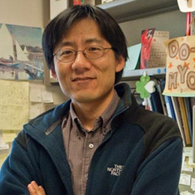
Bio
The Kim Lab is interested in understanding the molecular networks that coordinate nutrient metabolism and cell growth. How cells assess nutrient- or energy states and relay this information into appropriate decisions on growth is poorly understood. Coordinate regulation of nutrient metabolism and cell growth is of fundamental importance, and many human diseases, such as cancer, diabetes, and developmental disorders, are affected by alterations in this process.
Research Summary
mTOR signaling network
Our research is focused on the mTOR signaling network that plays a crucial role in controlling cell growth in response to nutrient levels and growth factors. mTOR binds several proteins to form two distinct protein complexes. mTORC1 (mTOR complex 1) contains raptor (KOG1 ortholog), Gbl/mLst8, PRAS40 and DEPTOR, whereas mTORC2 (mTOR complex 2) conatins rapamycin insensitive companion of mTOR (rictor) (Avo3 ortholog), GbL/mLst8, Sin1 (Avo1 ortholog), PRR5/protor and DEPTOR. In spite of considerable efforts, it has not been possible to obtain a clear understanding of the molecular mechanisms by which the mTOR network is regulates by nutrient- and growth factor-signals. Utilizing novel molecular biology and biochemical tool as well as a variety of structural approaches, we identify novel components and connectivity in the network and determine biological functions and signaling specificity thereof. Our recent studies have led us to identify PRAS40 and PRR5 as key components of mTORC1 and mTORC2. We investigate the functions of these newly-identified components in the regulation of cell growth and metabolism with links to human diseases such as cancer and diabetes. We anticipate this study will advance our understanding of the molecular bases underlying the coordinate regulation between metabolism and growth during animal development and the pathogenesis of metabolic diseases such as cancer and diabetes.
Autophagy pathway
As a crucial pathway downstream of mTOR, autophagy (cellular self-eating) plays an important role for metabolic homeostasis and cellular survival. Autophagy is an evolutionarily-conserved process through which cytoplasm, organelles, or long-lived proteins or protein aggregates are sequested in a double-membrane structures and subsequently degraded in lysosomes. Through destruction of cellular organelles and proteins, autophagy provides energy for starved cells or allows for balanced regulation between biogenesis and degradation of cellular structures, thereby playing important roles in growth, survival, differentiation, and development. Dys-regulation of autophagy is associated with many human diseases including cancer, myopathies, innate immunity, and neurodegenerative diseases such as Parkinson's and Huntington's diseases. Autophagy is induced when cells are starved of nutrients or mTOR is inhibited. Our recent study revealed that ULK1 and ULK1 protein kinases play key roles in autophagy induction in mammalian cells. We determined that mTORC1 phosphorylates ULK1 and ULK2 to inhibit the kinase functions. Later, studies from other groups identified that AMPK phosphorylates and positively regulates ULK1. We investigate how mTORC1 and AMPK regulate ULK functions with focus on its phosphorylation and its interaction with Atg13 and FIP200. We also study the shared and distinct functions of ULK1 and ULK2 in autophagy and non-autophagy processes.
mTORC1-regulated immunoproteasome assembly pathway
Protein stress, such as misfolded protein aggregates and oxidized proteins, occurs in many human diseases, including cancer, diabetes, neurodegeneration, and age-related pathol- ogies. One major source of protein stress originates when the ribosomal protein synthesis is upregulated with a consequential reduction of translational fidelity, producing defective ribosomal products. Prevalent in many cell growth disorders, such cancer and tuberous sclerosis complex (TSC), mutations of PTEN (phosphatase and tensin homolog), RAS, TSC1 (tuberous sclerosis complex 1), or TSC2 commonly enhance the activity of mTORC1 (mechanistic target of rapamycin complex 1). Hyper-activation of mTORC1, the central regulator of the ribosomal protein synthesis, can reduce translational fidelity as a result of increased rates of ribosomal elongation. The resulting aberrant proteins, if not properly removed, can cause cell death or contribute to human diseases.Our recent study showed that mTORC1 promotes the formation of immunoproteasomes for efficient turnover of defective proteins and cell survival. mTORC1 sequesters precursors of immunoproteasome beta subunits via PRAS40. When activated, mTORC1 phosphorylates PRAS40 to enhance protein synthesis and simultaneously to facilitate the assembly of the beta subunits for forming immunoproteasomes. Consequently, the PRAS40 phosphorylations play crucial roles in clearing aberrant proteins that accumulate due to mTORC1 activation. Mutations of RAS, PTEN, and TSC1, which cause mTORC1 hyperactivation, enhance immunoproteasome formation in cells and tissues. Those mutations increase cellular dependence on immunoproteasomes for stress response and survival. The results have defined a mechanism by which mTORC1 couples elevated protein synthesis with immunoproteasome biogenesis to protect cells against protein stress. The novel pathway (mTORC1-immunoproteasome) may regulate many other cellular and physiological processes, and its disturbance may lead to human diseases associated with cancer, neurodegenerative diseases, and autoimmune diseases.
Contact
Address
7-132 MCB420 Washington Ave SE
Minneapolis, MN 55455


Bio
Dr. Nichole (Nikki) Klatt is a Professor and the Director of Outcomes and Precision Medicine Research in the Department of Surgery, University of Minnesota. Prior to this she was an associate professor, vice chair of research, and the Adrienne Arsht Endowed Chair in Pediatric clinical research in the Department of Pediatrics, Miller School of Medicine, and Sylvester Cancer Center of University of Miami from 2018-2020. Dr. Klatt was also an associate professor in the Department of Pharmaceutics at the University of Washington from 2012- 2018. Dr. Klatt received her PhD from Emory University in Immunology and Molecular Pathogenesis in 2009, and was also a visiting PhD candidate at the University of Pennsylvania from 2006-2008. Dr. Klatt performed her postdoctoral research in the Laboratory of Molecular Microbiology at the National Institutes of Health from 2009-2012. The goal of Dr. Klatt's research laboratory is to understand mechanisms of microbiome, mucosal dysfunction and drug metabolism to improve human health.
Research Summary
The goal of Dr. Klatt's research laboratory is to understand mechanisms of the microbiome, mucosal dysfunction and drug metabolism to improve human health.
Selected Publications
Selected Presentations


Research Summary
Immunity to influenza virus infectionsInfluenza A virus (IAV) represents a major global health burden. Despite yearly vaccinations the virus is able to escape seasonal immunity requiring yearly vaccination and the threat of novel pandemics loom. Therefore continued understanding of the host-pathogen interactions and protective immune responses are critical for broadly protective vaccine development. Our overall research goal is to address fundamental questions in virology and viral immunology that have been difficult to dissect using conventional approaches. We utilized host-derived microRNAs, small non-coding RNA capable of mediating silencing of mRNA, to restrict the natural tropism of IAV. This allows for previously unavailable insights into immune responses to the virus. Additionally, we generate novel reporter viruses to further define the relationship between cellular tropism and immunity as well as to determine infected cell fate. A more comprehensive understanding of virus infection requirements that dictate immunity will be critical for the design of next generation vaccines and therapeutics
Contact
Address
1-117 MRF689 23rd Ave SE
Minneapoli, MN 55455


Bio
Michael Lipscomb is an Associate Professor in the Department of Pharmacology and the Associate Dean for Graduate Education in the Medical School. He received his B.S. degree from University of California Los Angeles with a degree in Microbiology, Immunology and Molecular Genetics prior to receiving a Ph.D. degree in Immunology from the University of Pittsburgh. His postdoctoral training was at University of Pennsylvania and Children's Hospital of Philadelphia. Dr. Lipscomb joined the Department of Pharmacology in 2022, previously a faculty member at Howard University, and is affiliated with the Center for Immunology.
Research Summary
Research in the Lipscomb Lab employs both basic and translational approaches to delineate the immunoregulatory networks that govern antigen presenting myeloid cell development and function. Investigations focus on defining the roles of novel genes that regulate dendritic cells (DC), monocyte and macrophage intracellular signaling events that include modulation of Ca2+ and cyclic nucleotides levels, activation/inactivation of protein kinases and post-translational modifications that alter downstream functional responses. In a related series of studies, the lab investigates how antigen presenting myeloid cells contribute to autoimmune disorders. Inflammatory regulating genes present in the MHC class III locus within the myeloid groups are studied to determine their collective contribution to initiating, sustaining and directing autoimmune pathologies. Lastly, the Lipscomb laboratory employs biomaterial compounds seeded with immune cells to generate new organoid structures as an innovative means of tissue restoration for alleviating immunodeficiency disorders.
Contact
Address
3-108 Nils Hasselmo Hall312 Church St SE
Minneapolis, MN 55455-0215
Bio
Administrator Info
Name: Admin
Phone: 612-624-9996
Fax: 612-625-4410
Email: idimdivision@umn.edu
Mail: Microbiology Research Building, 1st floor mail room, MMC 2821, 689 23rd Ave S.E., Minneapolis, MN 55455
Summary
Research in our neurovirology laboratory specifically investigates the role of CNS-infiltrating peripheral immune cells in driving chronic activation of brain-resident glial cells following viral infection. We are currently investigating the role of CD19(-)CD38(+)CD138(+) plasma cells and antiviral antibodies persisting within the CNS during chronic herpesvirus brain infection. We are also applying our viral brain infection models to study experimental immune reconstitution disease of the CNS (CNS-IRD) using T-cell repopulation of lymphopenic hosts (MAIDS animals) harboring HSV brain infection. As well as in the brain, dysregulated chronic immune activation and immune cell infiltration likely promote analogous nerve damage and neurotoxicity within the lumbar spinal cord (LSC) and dorsal root ganglia (DRG). These findings have led us to new studies on the neuropathogenesis of LP-BM5 retrovirus infection (i.e., MAIDS)-induced peripheral neuropathy.
Research Summary
Our research laboratory studies the pathogenesis of viral brain infection in mice. We work in the field of neurovirology; which is at the interface of virology, immunology, and neuroscience. We are a biomedical research laboratory, working on modeling disease processes. We currently have two projects funded through individual R01 grants from the National Institutes of Health. In the first project, funded through NINDS, we are investigating whether recall immune responses from virus-specific, brain-resident memory T cells (bTRM) activate brain-resident glia and induce production of neurotoxic mediators. Our goal is to determine whether adaptive recall responses to viral Ag trigger tissue-wide innate immune responses from reactive glia and promote inflammation-induced synaptic damage, neurotoxicity, and long-term neurocognitive impairment. In our second project, which is funded through the NIMH, we are trying to determine whether glial cells are viable cellular targets for immunotherapy. Microglia are the main reservoir for HIV-1 within the brain and potential exists for negative immune checkpoint blockade immunotherapies to purge viral reservoirs. There is currently a great deal of research interest in using checkpoint inhibitors to purge HIV in “cure” strategies. The vast majority of these trials target the PD-1: PD-L1 pathway. In this project, we are investigating cytolytic responses of CD8+ T lymphocytes against primary microglia loaded with viral peptide epitopes to determine whether immune checkpoint blockade targeting this pathway may be beneficial in clearing viral brain reservoirs.
Education
Fellowships, Residencies, and Visiting Engagements
Bio
Shawn Mahmud is a Pediatric Rheumatologist who completed MD/PhD (MSTP), residency, and fellowship training at the University of Minnesota. He provides care to children with autoimmune and autoinflammatory diseases including juvenile idiopathic arthritis, systemic lupus erythematosus, dermatomyositis, mixed connective tissue disease, chronic noninfectious osteitis, vasculitis, and others. He has a long-standing interest in immunology, specifically CD4+ T cell biology. During his graduate training, Dr. Mahmud made contributions to our understanding of how regulatory T cells develop in the thymus. His current focus is in developing peptide:MHCII tetramer-based approaches to identify, track, and phenotype important antigen-specific populations of CD4+ T cells in human disease.
Education
Fellowships, Residencies, and Visiting Engagements
Licensures and Certifications
Contact
Address
Pediatric Rheumatology, Allergy, & ImmunologyAcademic Office Building
2450 Riverside Ave S AO-10
Minneapolis, MN 55454

Research Summary
T cell migration, differentiation, and memory development in response to infectionsMy laboratory studies CD8 and CD4 T cell responses to a variety of viral and bacterial infections to help understand the development of immunological protection from re-infection. We observe and manipulate pathogen specific T cell responses over time by using MHC tetramers, adoptive transfer of transgenic T cells, fluorescence flow cytometry and sorting, and gene microarry analysis. Upon activation in lymphoid tissue, rare pathogen-specific naïve T cells proliferate, acquire effector functions, disseminate throughout the organism, and contribute to the eradication of pathogens. In situations where antigen is successfully eliminated, most effector T cells die by apoptosis. However, a fraction of effector T cells escape death and differentiate into long-lived memory T cells that contribute to protective immunity. We are currently dedicated to elucidating the developmental cues that govern T cell migration to different anatomical locations, commitment to the memory lineage, and the contribution of memory T cell differentiation state and location to protection from re-infection. Memory T cells that reside at common portals of pathogen entry or infection, especially the intestinal mucosa, are of particular interest. By understanding these issues, we hope to contribute to the development of better vaccination strategies, and are currently focused on informing development of a protective HIV vaccine.
Contact
Address
2-182 MBB2101 6th Street SE
Minneapolis, MN 55414


Bio
Dr. Arthur J. Matas is a Professor of Surgery at the University of Minnesota – Twin Cities. Dr. Matas earned his medical degree in 1972 at the University of Manitoba, in Winnipeg, Canada. He did his surgical residency and transplantation fellowship at the University of Minnesota Hospitals, where he was actively involved in clinical and laboratory research.Dr. Matas has authored or coauthored numerous articles and book chapters and his research has been presented at numerous national and international meetings. Dr. Matas is a long-standing member of many surgical and transplant-related societies including the American Society of Transplant Surgeons (of which he is Past President), the American Society of Transplantation, The Transplantation Society, the American Society of Nephrology, the American Surgical Association, and the American College of Surgeons.
Research Summary
My entire professional career has been in academic medicine, with a major focus on the identifying risk factors for poor long-term kidney transplant recipient outcomes; studies to improve short- and long-term recipient outcomes; and study of long-term living kidney donor outcomes.
Clinical Summary
Kidney Transplant, Living Donor Transplant
Education
Fellowships, Residencies, and Visiting Engagements
Honors and Recognition
Professional Memberships
Contact
Address
420 Delaware St SEPWB 11-200
Minneapolis, MN 55455
Administrative Contact
Carly Ryan | 612-625-5609 | ryan1025@umn.edu

Bio
Administrator Info
Name: Amirah Muwahid
Phone: 612-626-4024
Lab Phone: 612-626-4217
Email: muwah012@umn.edu
Mail: 420 Delaware Street SE
MMC 806
Minneapolis, MN 55455
Summary
Dr. Miller has been interested in NK cell biology, NK cell development, the acquisition of NK cell receptors and seamless translation into clinical trials throughout his entire academic career. Currently, the lab is focused on mechanisms which determine the enhanced function seen with CMV induced adaptive NK cells, facilitating immune synapses with IL-15 containing Trispecific Killer Engagers (TriKEs), IL-15 biology, NK cell killer immunoglobulin receptor (KIR) acquisition and function (NK cell education), and developing NK cell therapeutics.
Throughout his career at the University of Minnesota, he has mentored faculty and delivered hundreds of NK cells products to patients with cancer. His team has identified unique NKC2C+ NK cell repertoires exhibit a methylation signature of CD8+ T cells with properties of immune memory. Adaptive NK cells are distinctly different from canonical NK cells and signal through CD16 using a dominant CD3? signal by downregulation of FcR?R1?. Adaptive NK cells are better primed for killing, cytokine production, ADCC and exhibit unique metabolic signatures that enhance their survival. He has developed state-of-the-art functional readouts to study NK cells from the laboratory and the clinic based on high resolution testing. He was the first to report that haploidentical allogeneic human NK cells can persist and expand for up to one month after adoptive transfer. Based on these studies a significant part of his effort is trying to understand how to exploit NK cells for therapeutic purposes against infection and cancer and how to improve outcomes from allogeneic hematopoietic cell transplantation.
Clinically, he has developed first-in-human trials using allogeneic donor NK cells, rhIL-15, IL-15/IL-15R?-Fc (ALT-803, now called N-803), an NK cell TriKE that engages NK cells and AML targets that costimulates NK cells with an IL-15 linker. Dr. Miller's experience and translational expertise has supported a transition from individual related donor NK cell products to induced pluripotent stem cell (iPSC) derived NK cells. Advantages of using this off-the-shelf platform include the flexibility of multiple gene edits, immediate cryopreserved product availability, multi-doing strategies and combinatorial therapy with targeted agents to enhance NK cell function.
Dr. Miller is devoted to team science and mentoring. He has supervised > 400 NK cell products and sponsored >10 INDs and his team has studied >4000 transplant patients. Dr. Miller's experience, NIH grants and translational expertise provides a rich environment for training.
Research Summary
Natural Killer (NK) Cell Development
Throughout his academic career Dr. Miller has been interested in NK cell biology. His laboratory is focused on understanding the mechanisms of NK cell development and determining how NK cell killer immunoglobulin receptor (KIR) acquisition affects NK cell function through a process referred to as NK cell education or licensing. Most recently, he has been exploring the role of cytomegalovirus (CMV) reactivation after hematopoietic cell transplantation in enhancing NK cell reconstitution and function. CMV is the only virus known to induce the development of ”adaptive” NK cells with memory properties which are long lived and exhibit enhanced responses to subsequent exposures. Dr. Miller and his team have identified these adaptive NK cells in humans and determined that they have a methylation signature remarkably similar to that of CD8+ T cells. His long-term goal is to translate these novel findings into better NK-cell based immunotherapies to treat cancer without the morbidity of CMV infection.
Targeted Immunotherapy to Treat Human Cancer
Dr. Miller was the first to report that related donor haploidentical allogeneic NK cells can expand after adoptive transfer and induce remission in patients with refractory leukemia. Building on this landmark study, he spends significant effort developing novel methods to exploit NK cells therapeutically to treat infections, to cure cancer and to improve outcomes from allogeneic hematopoietic cell transplantation (HCT). He also leads a focused effort to understand the association of KIR immunogenetics with protection against relapse after allogeneic HCT. Dr. Miller and his team have demonstrated that transplants from donors with favorable KIR genes protect against relapse of acute myeloid leukemia after unrelated donor HCT. He is currently testing novel strategies to activate NK cells in vivo (using novel proteins such as interleukin-15) and to create antigen-specific NK cells with Bispecific Killer Engagers (BiKEs), which are proteins that facilitate targeting of tumor antigens by NK cells.
Current Discovery & Research Themes in the Miller Laboratory:
- NK cells and their receptors after hematopoietic cell transplantation
- CMV induced adaptive NK cells exhibit enhanced function through CD16
- Induction of NK cells antigen specificity through CD16 targeting
- Preserving and enhancing NK cell function through antibody dependent cellular cytotoxicy (ADCC)
Clinical Summary
Bone marrow transplant; Cancer immunotherapy
Education
Honors and Recognition
Professional Memberships


Bio
Administrator Info
Name: Drew Keup
Email: keupx013@umn.edu
Summary
Dr. Jerry A. Molitor is an Associate Professor of Medicine in the Division of Rheumatic and Autoimmune Diseases at the University of Minnesota Medical School. Dr. Molitor earned his PhD in Microbiology and Immunology at Duke University, where he helped develop evidence of a family of NF-kappa B nuclear transcription factors in T lymphocytes. He completed his MD at the University Of Iowa College Of Medicine. Dr. Molitor completed his Internal Medicine Internship and Residency, as well as a Rheumatology Fellowship, at the University of Washington. Dr. Molitor conducted numerous clinical and translational research projects through the Benaroya Research Institute at Virginia Mason, where he was Director of the Arthritis Clinical Research Unit from 2001-2007, and Associate Director of Clinical Research from 2004-2007. He joined the University of Minnesota in 2007, where he heads the multidisciplinary Scleroderma clinic. He has participated in the Scleroderma GWAS, and in multiples studies of Systemic Sclerosis biomarkers and natural history, as well as in various aspects of drug development. Dr. Molitor has clinical research interests in Early Rheumatoid Arthritis and Systemic Sclerosis pathogenesis and disease prevention, and has been a consultant or investigator in multiple trials of candidate therapies for these conditions. His translational research focuses on understanding the interplay between impaired immune responses to infections and associated autoimmunity. He leads current studies examining this phenomenon in individuals with periodontal disease and the development of Rheumatoid Arthritis–associated autoantibodies.
Clinical Summary
Rheumatology; Early rheumatoid arthritis; Scleroderma; Systemic Sclerosis; Polymyocitis/Dermatomyositis
