University of Minnesota Developing Compact and Portable MRI Scanner
Magnetic resonance imaging (MRI) is a powerful diagnostic tool that is out of reach for many clinics and smaller hospitals. That may be about to change. A shift in thinking about magnetic fields for diagnostic imaging recently resulted in a portable MRI scanner at the University of Minnesota Medical School. The U of M Medical School has received National Institutes of Health (NIH) grants that allow them to work on an international, multi-institutional project to continue to test and develop a new magnetic resonance imaging (MRI) scanner that is compact and transportable.
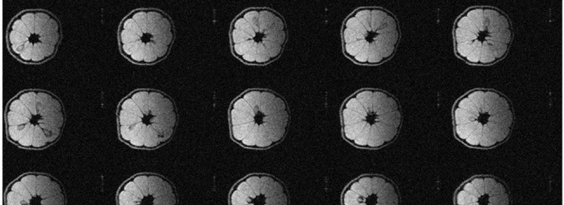
Michael Garwood, PhD, a professor and associate director of the Center for Magnetic Resonance Research at the U of M Medical School, and his team have been scanning lemons and water bottles with the device they created, and the images are as clear as those from fixed MRIs that use extremely heavy magnets.
“Over the past eight years, with funding from the NIH BRAIN Initiative and other sources, we have created MRI technology that allowed us to develop a prototype brain MRI scanner that promises to expand existing MRI capabilities and accessibility,” says Dr. Garwood.
Collaboration on this device among universities, countries and organizations has been seamless, with collaborators including imaging experts from Harvard, Yale and Columbia Universities; electronic component experts in Brazil; and electrical superconductor experts in New Zealand.
Human trials could begin as early as this summer if the scanner passes all the necessary safety testing. Their goal is to expand access to people in more rural areas, where many people have to travel quite far to receive any kind of advanced diagnostic imaging.
MRIs have existed for nearly half a century. They use extremely powerful, heavy magnets and radio waves to stimulate molecules in the body to produce precise images. MRI machines are expensive and require specialized facilities and trained professionals to operate and maintain them. These machines are so large it makes them difficult to install in some hospitals, making it hard to provide them in rural or resource-limited areas.
“The design of our new MRI head-only magnet, developed by the team at TE Herenga Waka in New Zealand, will allow studies of the human brain without a highly confining MRI magnet bore,” Dr. Garwood said. “Due to its small size and its light weight, this MRI scanner will be transportable for imaging brain function and structure almost anywhere, for almost anyone.”
In recent innovations, focusing on just one specific body part has been the way to go when developing these smaller MRI machines. Dr. Garwood and his team are focusing specifically on the brain, after first attempting to obtain funding to develop a compact MRI device for breast cancer screening.
In the Center for Magnetic Resonance Research, the team plans to use MRI techniques to create different types of images that are needed for various research purposes. Once they pass all of the required safety tests, they plan to collaborate with communities that have been historically known to have limited access to medical resources, such as Native American tribal communities, where they hope to conduct a pilot study of this technology in the field. The prototype looks like a throne with a half-dome magnet on top. It sits on wheels and can roll through doorways—as long as the 800-pound magnet is removed and moved separately.