Visible Heart Laboratories Reaches International Territories with Education
The Visible Heart® Laboratories at the University of Minnesota Medical School, is bridging the international gap; every year, over half a million people utilize their website. The mission of this group is to actively participate in educational outreach, and one of the biggest ways to do this is through their free-access website. Site visitors are able to view more than 5,000 videos and still images, including anatomy tutorials, in-depth descriptions of heart conditions and medical devices, such as MRIs and 3D modeling, which are all found under the Atlas tab.
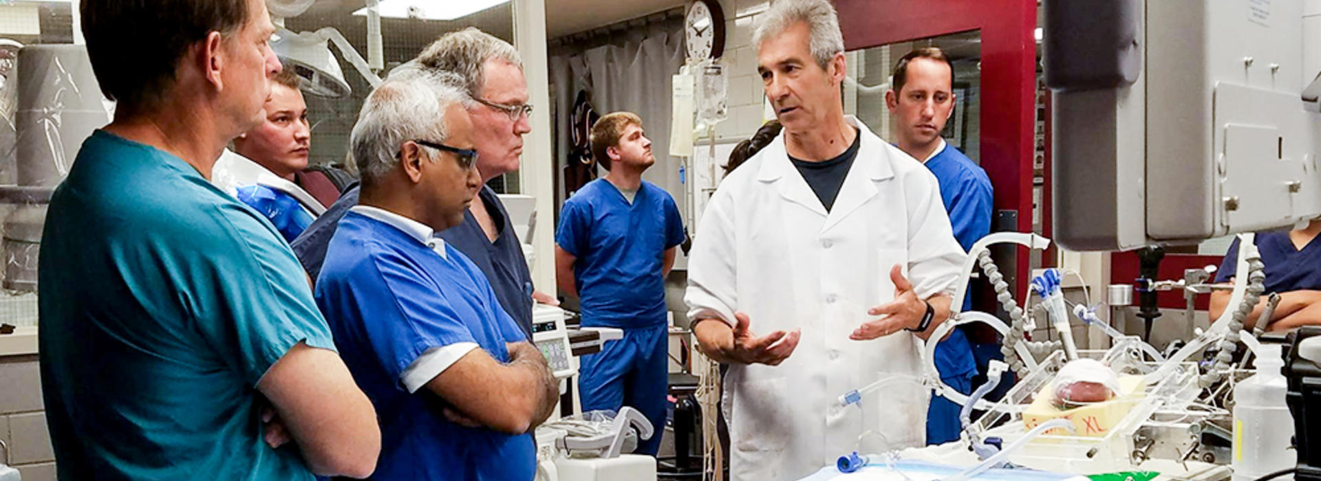
But, the educational outreach doesn’t end there. For over 20 years, the Visible Heart Laboratories has provided tours for students, including middle and high school groups, Medtronic employees and physicians around the globe. This research facility is considered by many as a premiere place to perform translational systems physiology research, which ranges from cellular and tissue studies to organ and whole body examinations. Physicians, fellows and students travel to the laboratories to take videos, test medical devices and grasp a deeper understanding of the anatomical features of the functioning human heart.
“On average, we interact directly with several thousand people as they visit our lab on an annual basis,” says Paul Iaizzo, PhD, a professor with the Visible Heart Laboratories.
This summer, Dr. Iaizzo will have worked within the Medical School for 30 years. In 1996, he started the Visible Heart Laboratories and has received consistent funding from Medtronics since 1997 for ongoing collaborative research. Dr. Iaizzo and his team routinely perform heart reanimations, develop novel ways to test medical devices and continue to provide new educational materials and programs related to the human heart.
If an organ donor’s heart is not viable for transplant, Dr. Iaizzo and his team may receive a call with the offer from LifeSource, an organ procurement organization, to use this heart for educational and research purposes with permission from the organ donor’s family. On rare occasions, if such a specimen arrives to the lab within four to six hours and there is a team available to reanimate it, then the action begins.
The team spends several hours collecting novel educational images that are added to the free-access Atlas website. Sometimes, they can uniquely visualize the functional anatomy of the human heart using a variety of imaging modalities. The first human heart reanimation was performed within the our Medical School in 2000, and since then Dr. Iaizzo and his team has reanimated 87 donor human hearts, with the last one being several weeks ago.
“We feel we owe it, to the organ donor and their family for providing us with this precious gift, to in turn, provide videos and images of this patient’s heart for all to learn from,” Dr. Iaizzo says. “No two human hearts are the same, just like humans having unique thumbprints. To me, the functional anatomy with the human heart is just beautiful.”
Dr. Iaizzo and his team also take their educational expertise on the road, bringing in heart models, videos and virtual reality systems to schools and science fairs with the mission to educate and spark interests within this area of medicine or science in general. Their annual calendar is full of educational outreach events, tours and visit requests, in which sometimes there are almost more requests than can be met. However, Dr. Iaizzo and his team try their best to make it work for everyone.
“One of the things I remind students, who are within the lab, is that you never know who will be moved, and if you impact even one student per group, then that tour was worth doing,” he says.
To learn more about the Visible Heart Laboratories, visit the website.