Tissue Clearing
Clearing
The Sturctural Circuits Core partners with the University Imaging Centers to integrate state-of-the-art tissue clearing methods and technologies to help investigators prepare, image, and analyze biologically intact tissues and organs in three dimensions. The group also works with researchers to optimize antibody labeling of cleared tissues with microwave-assisted methodology.
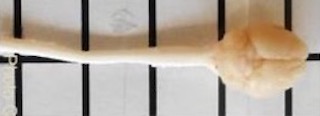
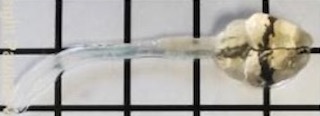
Tissue clearing brings a new dimension to histology services, where historically, tissue sectioning not only provides limited information of biological structures in its native form, but also proved to be a laborious and computationally intensive challenge in reconstruction from 2D to 3D. An assortment of tissue clearing methods has been developed in multiple laboratories within the last decade to make various tissues optically transparent by reducing light scattering intrinsic in tissues. This enables optimized whole-mount imaging on our specialized imaging systems, in particular, the Caliber ID RS-G4 Ribbon Scanning Confocal and the 3i Cleared Tissue Light Sheet (CTLS) microscope.
The UIC employs multiple tissue clearing approaches while primarily focusing on two: the PEGASOS organic-based and X-CLARITY hydrogel-based methods. PEGASOS and X-CLARITY are compatible with antibody and fluorescence staining, respectively, which allows for a comprehensive analysis of preserved internal structures.
z-series of neurons within the hippocampus of cleared mouse brain
X-CLARITY cleared mouse brain acquired using a 10x/0.5 NA Glyc objective on a Caliber ID RS-G4 Ribbon Scanning Confocal. This region shows the z-series of neurons within the hippocampus.
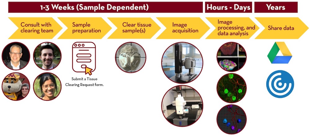
Tissue Clearing and Imaging Pipeline


Additional Information on Tissue Clearing
For additional information on tissue clearing see the UMN Imaging Center website.
Contact Information:
For general inquries about instrumentation, services, or projects please send a message at uic-staff@umn.edu. Once received, a member of the UIC team will followup to discuss your inquiry.